Japanese
English
- 有料閲覧
- Abstract 文献概要
- 1ページ目 Look Inside
- 参考文献 Reference
はじめに
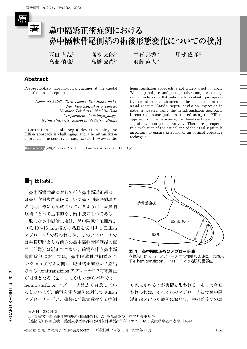
鼻中隔彎曲症に対して行う鼻中隔矯正術は,耳鼻咽喉科専門研修において鼻・副鼻腔領域での到達目標にも記載されているように,耳鼻咽喉科にとって基本的な手術手技の1つである。一般的な鼻中隔矯正術は,鼻中隔軟骨尾側端より約10〜15mm後方の粘膜を切開するKillianアプローチ1)で行われるが,このアプローチでは粘膜切開よりも前方の鼻中隔軟骨尾側端の彎曲(前彎)は矯正できない。前彎を伴う鼻中隔彎曲症例に対しては,鼻中隔軟骨尾側端から2〜3mm後方を切開し,尾側端を前方から露出させるhemitransfixionアプローチ2)で前彎矯正が可能となる(図1)。しかしながら本邦では,hemitransfixionアプローチは広く普及しているとはいえず,前彎を伴う症例に対してKillianアプローチを行い,術後に前彎が残存する症例も散見されるのが実情と思われる。そこで今回われわれは,それぞれのアプローチ法で鼻中隔矯正術を行った症例において,手術前後での鼻中隔軟骨尾側端の形態変化を明らかにするために,CTを用いて検討を行ったので報告する。
Correction of caudal septal deviation using the Killian approach is challenging, and a hemitransfixion approach is necessary in such cases. However, the hemitransfixion approach is not widely used in Japan. We compared pre- and postoperative computed tomography findings in 104 patients to evaluate postoperative morphological changes at the caudal end of the nasal septum. Caudal septal deviation improved in patients treated using the hemitransfixion approach. In contrast, some patients treated using the Killian approach showed worsening or developed new caudal septal deviation postoperatively. Therefore, preoperative evaluation of the caudal end of the nasal septum is important to ensure selection of an optimal operative technique.

Copyright © 2022, Igaku-Shoin Ltd. All rights reserved.


