Japanese
English
臨床報告 カラー臨床報告
硝子体手術と内境界膜剥離を行った嚢胞様糖尿病黄斑浮腫3眼の光干渉断層計による評価
Optical coherence tomographic features of three eyes of diabetic cystoid macular edema treated by vitrectomy and inner-limiting-membrane maculorhexis
矢那瀬 淳一
1
,
荻野 誠周
1
,
栗原 秀行
1
Junichi Yanase
1
,
Nobuchika Ogino
1
,
Hideyuki Kurihara
1
1栗原眼科病院
1Kurihara Eye Hosp
pp.745-750
発行日 1999年4月15日
Published Date 1999/4/15
DOI https://doi.org/10.11477/mf.1410906324
- 有料閲覧
- Abstract 文献概要
- 1ページ目 Look Inside
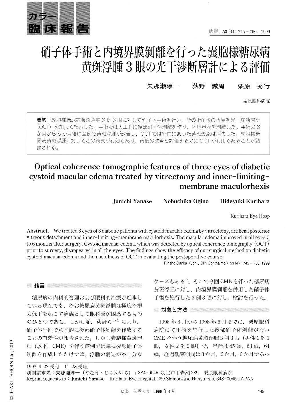
嚢胞様糖尿病黄斑浮腫3例3眼に対して硝子体手術を行い,その術前後の所見を光干渉断層計(OCT)を加えて検索した。手術では人工的に後部硝子体剥離を作り,内境界膜を剥離した。手術の3か月から6か月後に全例で黄斑浮腫が改善し,OCTでは術前にあった黄斑嚢胞は消失した。嚢胞様糖尿病黄斑浮腫に対してこの術式が有効であり,術後の改善を評価するのにOCTが有用であることが結論される。
We treated 3 eyes of 3 diabetic patients with cystoid macular edema by vitrectomy, artificial posterior vitreous detachment and inner-limiting-membrane maculorhexis. The macular edema improved in all eyes 3 to 6 months after surgery. Cystoid macular edema, which was detected by optical coherence tomography (OCT) prior to surgery, disappeared in all the eyes. The findings show the efficacy of our surgical method on diabetic cystoid macular edema and the usefulness of OCT in evaluating the postoperative course.

Copyright © 1999, Igaku-Shoin Ltd. All rights reserved.


