Japanese
English
- 有料閲覧
- Abstract 文献概要
- 1ページ目 Look Inside
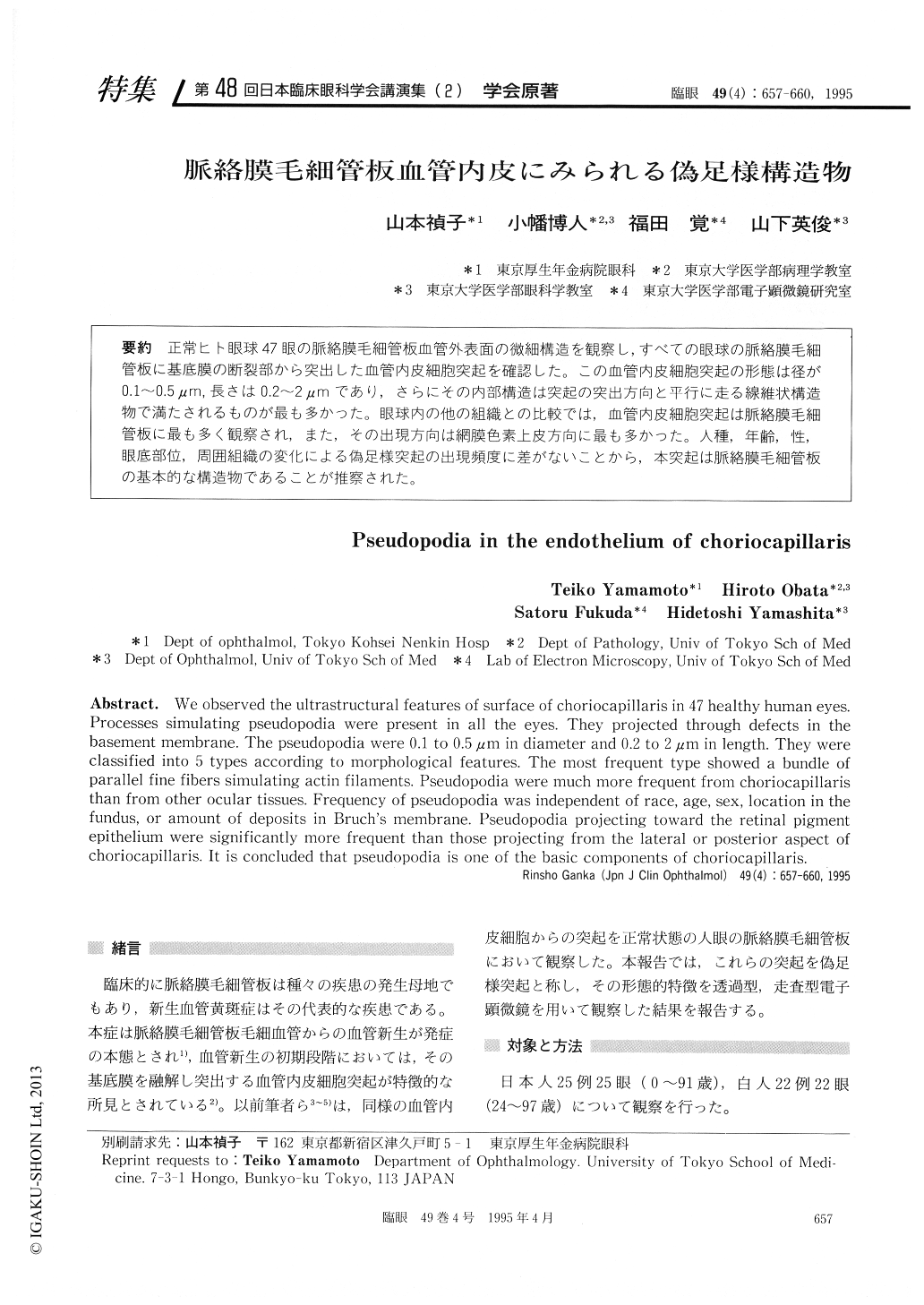
正常ヒト眼球47眼の脈絡膜毛細管板血管外表面の微細構造を観察し,すべての眼球の脈絡膜毛細管板に基底膜の断裂部から突出した血管内皮細胞突起を確認した。この血管内皮細胞突起の形態は径が0.1〜0.5μm,長さは0.2〜2μmであり,さらにその内部構造は突起の突出方向と平行に走る線維状構造物で満たされるものが最も多かった。眼球内の他の組織との比較では,血管内皮細胞突起は脈絡膜毛細管板に最も多く観察され,また,その出現方向は網膜色素上皮方向に最も多かった。人種,年齢,性,眼底部位,周囲組織の変化による偽足様突起の出現頻度に差がないことから,本突起は脈絡膜毛細管板の基本的な構造物であることが推察された。
We observed the ultrastructural features of surface of choriocapillaris in 47 healthy human eyes. Processes simulating pseudopodia were present in all the eyes. They projected through defects in the basement membrane. The pseudopodia were 0.1 to 0.5μm in diameter and 0.2 to 2μm in length. They were classified into 5 types according to morphological features. The most frequent type showed a bundle of parallel fine fibers simulating actin filaments. Pseudopodia were much more frequent from choriocapillaris than from other ocular tissues. Frequency of pseudopodia was independent of race, age, sex, location in the fundus, or amount of deposits in Bruch's membrane. Pseudopodia projecting toward the retinal pigment epithelium were significantly more frequent than those projecting from the lateral or posterior aspect of choriocapillaris. It is concluded that pseudopodia is one of the basic components of choriocapillaris.

Copyright © 1995, Igaku-Shoin Ltd. All rights reserved.


