Japanese
English
- 有料閲覧
- Abstract 文献概要
- 1ページ目 Look Inside
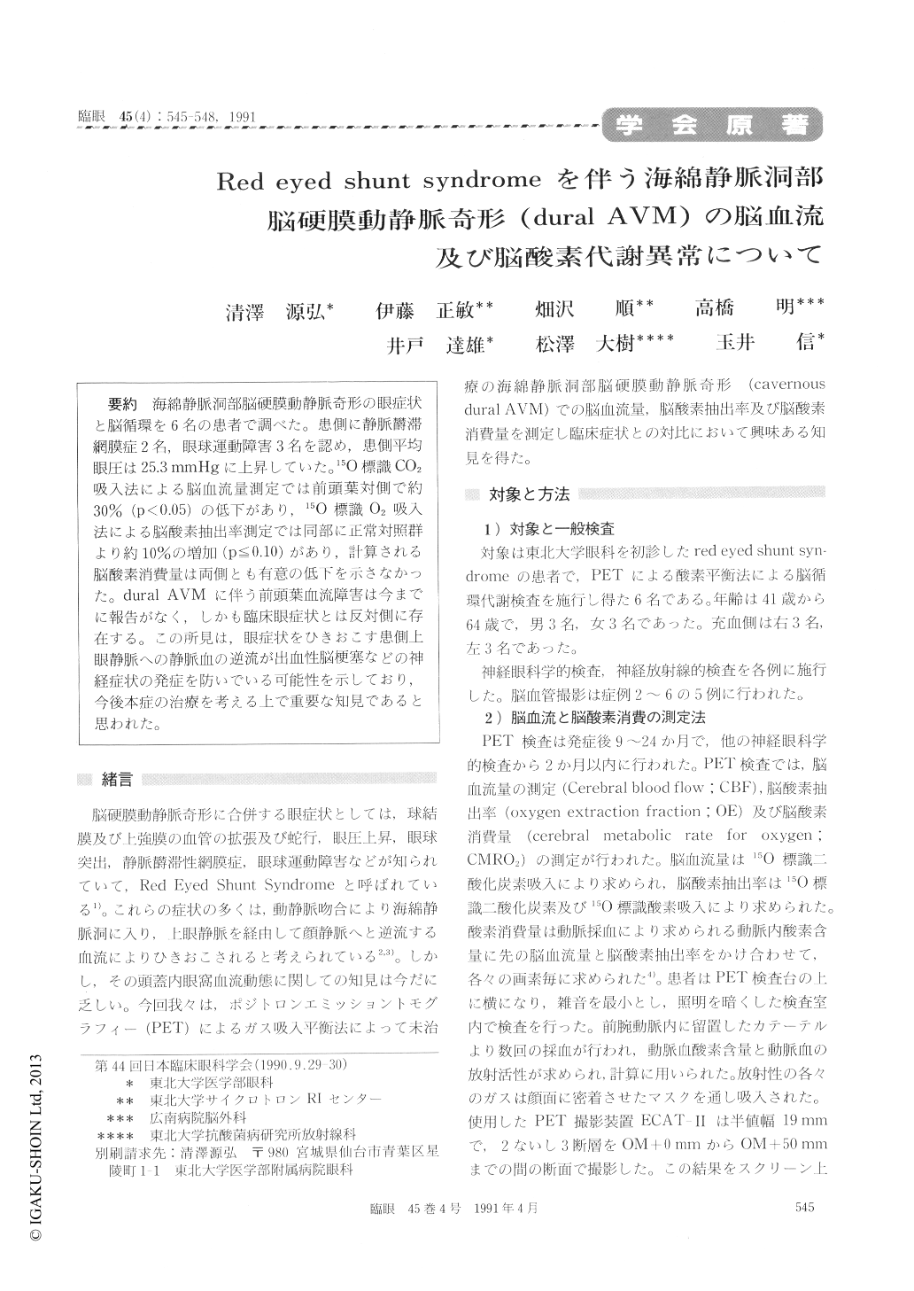
海綿静脈洞部脳硬膜動静脈奇形の眼症状と脳循環を6名の患者で調べた。患側に静脈欝滞網膜症2名,眼球運動障害3名を認め,患側平均眼圧は25.3mmHgに上昇していた。15O標識CO2吸入法による脳血流量測定では前頭葉対側で約30%(P<0.05)の低下があり,15O標識O2吸入法による脳酸素抽出率測定では同部に正常対照群より約10%の増加(P≦0.10)があり,計算される脳酸素消費量は両側とも有意の低下を示さなかった。dural AVMに伴う前頭葉血流障害は今までに報告がなく,しかも臨床眼症状とは反対側に存在する。この所見は,眼症状をひきおこす患側上眼静脈への静脈血の逆流が出血性脳梗塞などの神経症状の発症を防いでいる可能性を示しており,今後本症の治療を考える上で重要な知見であると思われた。
We studied ocular symptomes and cerebral blood flow and metabolism in 6 patients with cavernous dural arteriovenous malformations. Ophthal-mological examination revealed venous stasis retinopathy in 2 patients and disturbed ocular movement in 3. The mean intraocular pressure was increased to 25.3 mmHg. Positron emission tomo-graphy with 15O-labelled CO2 continuous inhalation revealed reduction of blood flow in the contralater-al frontal lobe by 30% (p< 0.05).
PET with 15O labelled CO2 continuous inhalation revealed increase in oxygen extraction rate by 10% (p≦0.10). Calculated cerebral motabolic rate for oxygen (CMRO2) showed no signilicant reduction.
This is the first report of dural AVM showing low cerebral blood flow in the contralateral frontal cortex. This finding suggests a possiblity to protect the brain by reversed flow of supra ophthalmic vein.

Copyright © 1991, Igaku-Shoin Ltd. All rights reserved.


