Japanese
English
- 有料閲覧
- Abstract 文献概要
- 1ページ目 Look Inside
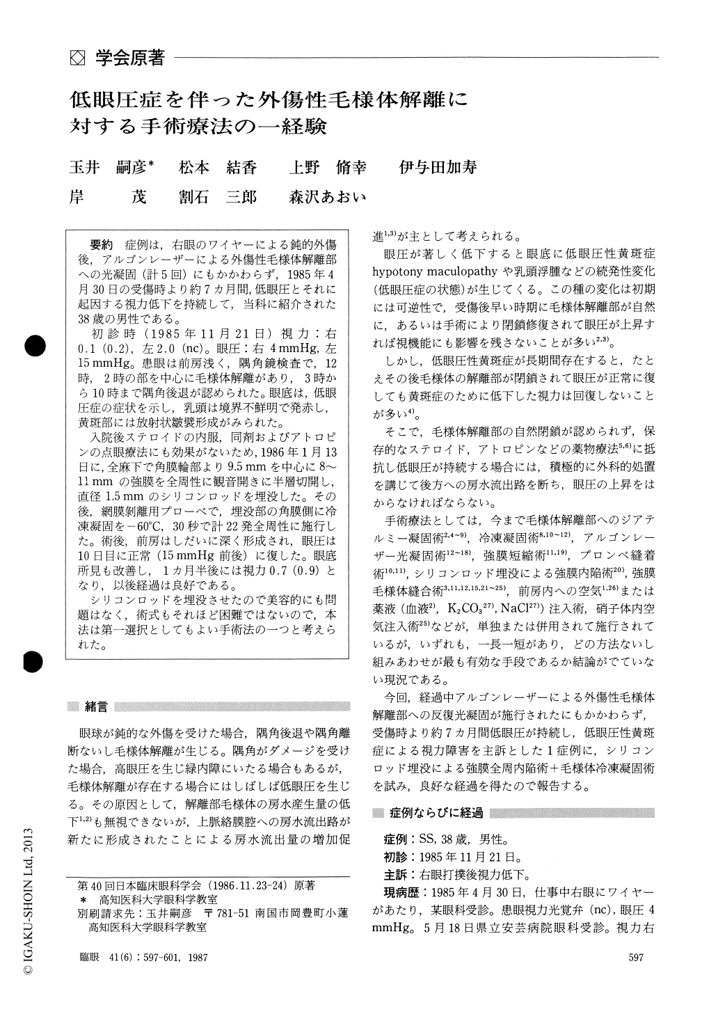
症例 は,右眼のワイヤーによる鈍的外傷後,アルゴンレーザーによる外傷性毛様体解離部への光凝固(計5回)にもかかわらず,1985年4月30日の受傷時より約7カ月間,低眼圧とそれに起因する視力低下を持続して,当科に紹介された38歳の男性である.
初診時(1985年11月21日)視力:右0.1(0.2),左2.0(nc).眼圧:右4mmHg,左15mmHg.患眼は前房浅く,隅角鏡検査で,12時,2時の部を中心に毛様体解離があり,3時から10時まで隅角後退が認められた.眼底は,低眼圧症の症状を示し,乳頭は境界不鮮明で発赤し,黄斑部には放射状皺襞形成がみられた.
入院後ステロイドの内服,同剤およびアトロピンの点眼療法にも効果がないため,1986年1月13日に,全麻下で角膜輪部より9.5mmを中心に8〜11mmの強膜を全周性に観音開きに半層切開し,直径1.5mmのシリコンロッドを埋没した.その後,網膜剥離用プローベで,埋没部の角膜側に冷凍凝固を−60℃,30秒で計22発全周性に施行した.術後,前房はしだいに深く形成され,眼圧は10日目に正常(15mmHg前後)に復した.眼底所見も改善し,1カ月半後には視力0.7(0.9)となり,以後経過は良好である.
シリコンロッドを埋没させたので美容的にも問題はなく,術式もそれほど困難ではないので,本法は第一選択としてもよい手術法の一つと考えられた.
A 38-year-old male developed ocular hypotony soon after blunt trauma with cyclodialysis in his right eye. Repeated argon laser photocoagulation to the chamber angle proved futile. He was referred to us 7 months after the injury. When seen first by us, the visual acuity was 0.2 and the intraocular pres-sure (IOP) was 4 mmHg in the right eye. The anterior chamber was extremely shallow. Hypotonymaculopathy was present.
We treated the patient with cryoapplication and scleral buckling by implanting a 1.5 mm silicone rod circumeferentially 9.5 mm posterior to the limbus.The IOP returned to the normal range ten days after surgery, followed by gradual improvement in hypotony maculopathy.
The present surgical method promises to be of value in the treatment of ocular hypotony due to traumatic cyclodialysis.
Rinsho Ganka (Jpn J Clin Ophthalmol) 41(6) : 597-601, 1987

Copyright © 1987, Igaku-Shoin Ltd. All rights reserved.


