Japanese
English
- 有料閲覧
- Abstract 文献概要
- 1ページ目 Look Inside
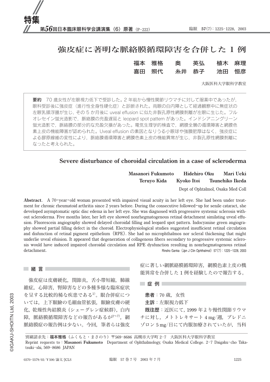
要約 70歳女性が左眼視力低下で受診した。2年前から慢性関節リウマチに対して服薬中であったが,眼科受診後に強皮症(進行性全身性硬化症)と診断された。両眼の白内障として経過観察中に無症状の左眼乳頭浮腫が生じ,その5か月後にuveal effusionに似た非裂孔原性網膜剝離が左眼に生じた。フルオレセイン蛍光造影で,脈絡膜の充盈遅延とleopard spot patternがあった。インドシアニングリーン蛍光造影で,脈絡膜の部分的な充盈欠損があった。電気生理学的検査で,網膜全層の循環障害と網膜色素上皮の機能障害が認められた。Uveal effusionの素因となりうる小眼球や強膜肥厚はなく,強皮症による膠原線維の変性により,脈絡膜循環障害と網膜色素上皮の機能異常が生じ,非裂孔原性網膜剝離になったと考えられた。
Abstract. A 70-year-old woman presented with impaired visual acuity in her left eye. She had been under treatment for chronic rheumatoid arthritis since 2 years before. During the consecutive followed-up for senile cataract,she developed asymptomatic optic disc edema in her left eye. She was diagnosed with progressive systemic sclerosis without scleroderma. Five months later,her left eye showed nonrhegmatogenous retinal detachment similating uveal effusion. Fluorescein angiography showed delayed choroidal filling and leopard spot pattern. Indocyanine green angiography showed partial filling defect in the choroid. Electrophysiological studies suggested insufficient retinal circulation and disfunction of retinal pigment epithelium(RPE). She had no microphthalmos nor scleral thickening that might underlie uveal efuision. It appeared that degeneration of collagenous fibers secondary to progressive systemic sclerosis would have induced impaired choroidal circulation and RPE dysfunction resulting in nonrhegmatogenous retinal detachment.

Copyright © 2003, Igaku-Shoin Ltd. All rights reserved.


