Japanese
English
- 有料閲覧
- Abstract 文献概要
- 1ページ目 Look Inside
- 参考文献 Reference
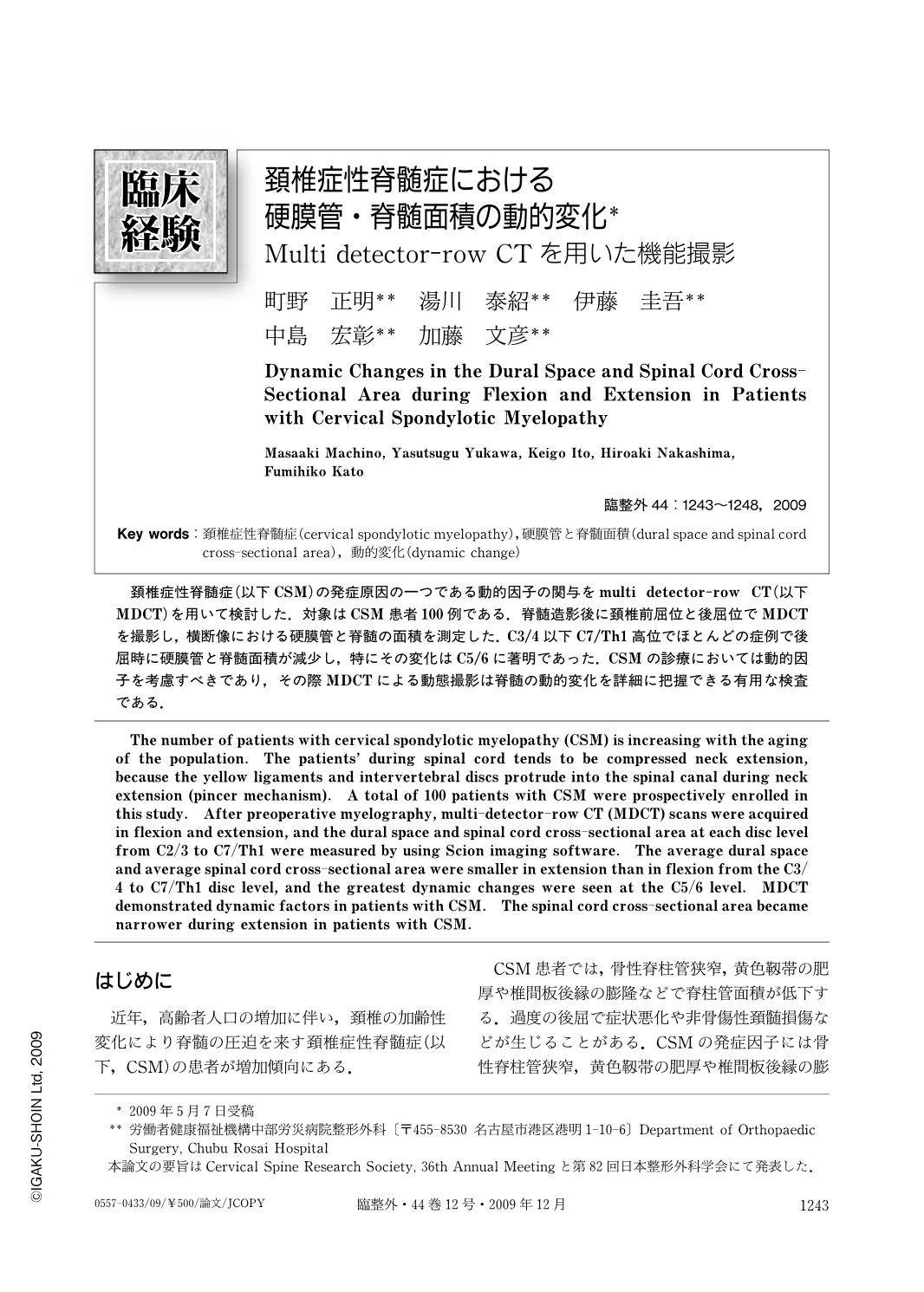
頚椎症性脊髄症(以下CSM)の発症原因の一つである動的因子の関与をmulti detector-row CT(以下MDCT)を用いて検討した.対象はCSM患者100例である.脊髄造影後に頚椎前屈位と後屈位でMDCTを撮影し,横断像における硬膜管と脊髄の面積を測定した.C3/4以下C7/Th1高位でほとんどの症例で後屈時に硬膜管と脊髄面積が減少し,特にその変化はC5/6に著明であった.CSMの診療においては動的因子を考慮すべきであり,その際MDCTによる動態撮影は脊髄の動的変化を詳細に把握できる有用な検査である.
The number of patients with cervical spondylotic myelopathy (CSM) is increasing with the aging of the population. The patients' during spinal cord tends to be compressed neck extension, because the yellow ligaments and intervertebral discs protrude into the spinal canal during neck extension (pincer mechanism). A total of 100 patients with CSM were prospectively enrolled in this study. After preoperative myelography, multi-detector-row CT (MDCT) scans were acquired in flexion and extension, and the dural space and spinal cord cross-sectional area at each disc level from C2/3 to C7/Th1 were measured by using Scion imaging software. The average dural space and average spinal cord cross-sectional area were smaller in extension than in flexion from the C3/4 to C7/Th1 disc level, and the greatest dynamic changes were seen at the C5/6 level. MDCT demonstrated dynamic factors in patients with CSM. The spinal cord cross-sectional area became narrower during extension in patients with CSM.

Copyright © 2009, Igaku-Shoin Ltd. All rights reserved.


