Japanese
English
- 有料閲覧
- Abstract 文献概要
- 1ページ目 Look Inside
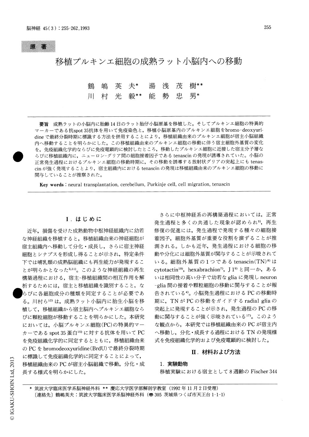
成熟ラットの小脳内に胎齢14日のラット胎仔小脳原基を移植した。そしてプルキンエ細胞の特異的マーカーである抗spot 35抗体を用いて免疫染色と,移植小脳原基内のプルキンエ細胞をbromo-deoxyuri—dineで最終分裂時期に標識する方法を併用することにより,移植組織由来のプルキンエ細胞が宿主小脳組織内へ移動することを明らかにした。この移植組織由来のプルキンエ細胞の移動に伴う宿主細胞外基質の変化を,免疫組織化学的ならびに免疫電顕的に検討したところ,移動したプルキンエ細胞に近接した宿主分子層ならびに移植組織内に,ニューロン‐グリア間の細胞接着因子であるtenascinの発現が誘導されていた。小脳の正常発生過程におけるプルキンエ細胞の移動時期に,その移動を誘導する放射状グリアの突起上にもtenas—cinが強く発現することより,宿主組織内におけるtenascinの発現は移植組織由来のプルキンエ細胞の移動に関与していることが推察された。
It is considered that cell adhesion molecules play important roles in the host-graft interaction during the reconstruction of the injured nervous system by neural transplantation. In this article, we report the expression of such molecules during the migration and differentiation of donor Purkinje cells in the adult rat cerebellum. Cerebellar primordium at the 14th day of gestation was transplanted into the adult rat cerebellum. Purkinje cells which had migrated from the grafted tissue into the host molecular layer were identified immunohisto-chemically with anti-spot 35 antibody, a specific marker for Purkinje cells in the cerebellum, as well as by labeling them with bromodeoxyuridine at their final mitotic period. In the grafted site, tran-sient expression of a neuron-glia cell adhesion molecule, tenascin, was detected immunohisto-chemically. This molecule was expressed in the host tissue adjacent to the migratory Purkinje cells as well as within the donor immature tissue. Tenascin was not detected in the host tissue far distant from the grafted tissue. In considering the expression of tenascin in the migratory process of Purkinje cells during cerebellar development, this molecule in-duced in the host tissue may be involved in the migration of donor Purkinje cells.

Copyright © 1993, Igaku-Shoin Ltd. All rights reserved.


