Japanese
English
- 有料閲覧
- Abstract 文献概要
- 1ページ目 Look Inside
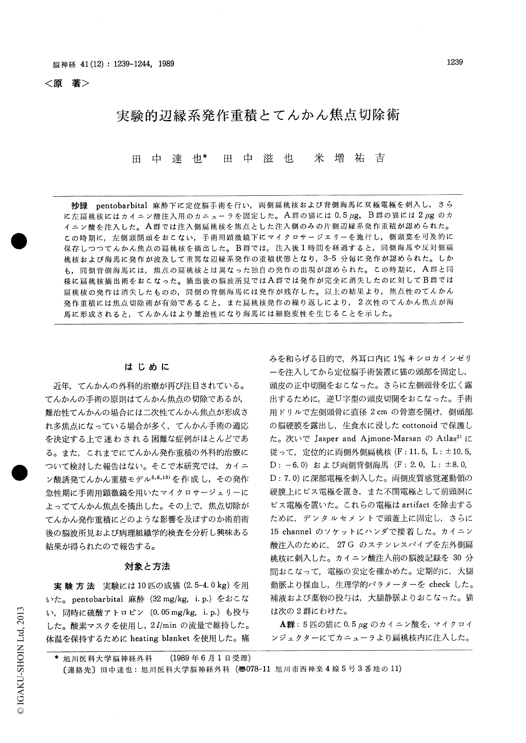
抄録 pentobarbital麻酔下に定位脳手術を行い,両側扁桃核および背側海馬に双極電極を刺入し,さらに左扁桃核にはカイニン酸注入用のカニューラを固定した。A群の猫には0.5μg, B群の猫には2μgのカイニン酸を注入した。A群では注入側扁桃核を焦点とした注入側のみの片側辺縁系発作重積が認められた。この時期に,左側頭開頭をおこない,手術用顕微鏡下にマイクロサージエリーを施行し,側頭葉を可及的に保存しつつてんかん焦点の扁桃核を摘出した。B群では,注入後1時間を経過すると,同側海馬や反対側扁桃核および海馬に発作が波及して重篤な辺縁系発作の重積状態となり,3-5分毎に発作が認められた。しかも,同側背側海馬には,焦点の扁桃核とは異なった独自の発作の出現が認められた。この時期に,A群と同様に扁桃核摘出術をおこなった。摘出後の脳波所見ではA群では発作が完全に消失したのに対してB群では扁桃核の発作は消失したものの,同側の背側海馬には発作が残存した。以上の結果より,焦点性のてんかん発作重積には焦点切除術が有効であること,また扁桃核発作の繰り返しにより,2次性のてんかん焦点が海馬に形成されると,てんかんはより難治性になり海馬には細胞変性を生じることを示した。
Status epilepticus is a neurological emergency and refractory one often resulted in neurological damage or death. Since the basic mechanisms of status epilepticus was not fully understood, a surgical treatment was not attempted until now. In the present study, a surgical resection of the epileptic focus was made in experimentally induced limbic status epilepticus and influences of the surgery upon status epilepticus was discussed.
Limbic status epilepticus was induced by means of kainic acid (KA) microinjection into unilateral amygdala in cats and effects of focus resection upon limbic seizure status were studied. Ten adult cats were stereotaxically operated on under pentobarbital anesthesia. Bipolar electrodes were placed in bilateral amygdala and hippocampus. An injection cannula, designed for kainic acid injection, was placed in the left amygdala. The cats were then divided into two groups. Group A (5 cats) received 0.5 μg of KA injection into the amygdala resulted in mild limbic status. Two of them were controls and 3 of them received amygdalotomy after induction of the limbic seizure status. Group B (5 cats) received 2.0/μg KA in-jection resulted in severe limbic status. Moreover, independent spontaneous seizure activities were observed in the ipsilateral hippocampus. Two of them were controls and 3 of them were operated on. After amygdalotomy, limbic seizure stopped in the operated cats of Group A. In the operated cats of Group B, repeated seizures in the epilepto-genic focus (amygdala) was completely suppressed, however, spontaneous seizures of the ipsilateral hippocampus persisted even after the surgery.
Histopathological study confirmed that the left amygdala, primary epileptogenic focus, was com-pletely resected in operated cats. Histopatholo-gical changes were not observed in Group A.
On the contrary, mild pyknosis was observed in the CA 3 portion of the pyramidal cell layer of the ipsilateral hippocampus in Group B. The present study revealed that surgical resection of the epileptogenic focus was effective for the treat-ment of limbic seizure status epilepticus, when the focus was restricted and secondarily epilepto-genic focus was not observed. But, the surgery was no more effective for the refractory limbicstatus epilepticus. In the histopathological study, neuronal cell damage was already observed in the ipsilateral hippocampus within 8 hours after KA injection into the amygdala. The results suggested that prolonged limbic status epilepticus could cause hippocampal damage due to hippocampal vulner-ability to the seizures.

Copyright © 1989, Igaku-Shoin Ltd. All rights reserved.


