Japanese
English
- 有料閲覧
- Abstract 文献概要
- 1ページ目 Look Inside
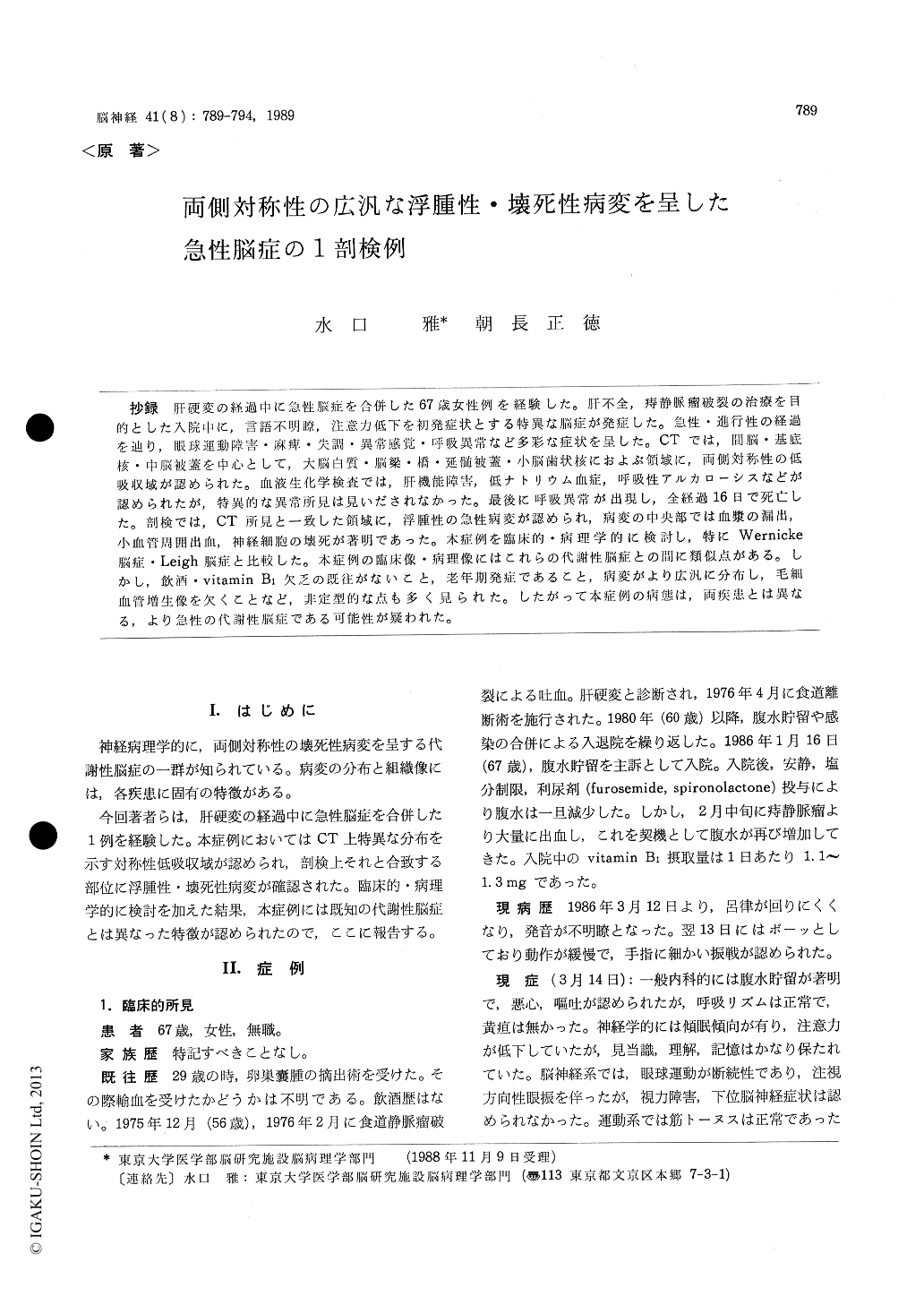
抄録 肝硬変の経過中に急性脳症を合併した67歳女性例を経験した。肝不全,痔静脈瘤破裂の治療を目的とした入院中に,言語不明瞭,注意力低下を初発症状とする特異な脳症が発症した。急性・進行性の経過を辿り,眼球運動障害・麻痺・失調・異常感覚・呼吸異常など多彩な症状を呈した。CTでは,間脳・基底核・中脳被蓋を中心として,大脳白質・脳梁・橋・延髄被蓋・小脳歯状核におよぶ領域に,両側対称性の低吸収域が認められた。血液生化学検査では,肝機能障害,低ナトリウム血症,呼吸性アルカローシスなどが認められたが,特異的な異常所見は見いだされなかった。最後に呼吸異常が出現し,全経過16日で死亡した。剖検では,CT所見と一致した領域に,浮腫性の急性病変が認められ,病変の中央部では血漿の漏出,小血管周囲出血,神経細胞の壊死が著明であった。本症例を臨床的・病理学的に検討し,特にWernicke脳症・Leigh脳症と比較した。本症例の臨床像・病理像にはこれらの代謝性脳症との間に類似点がある。しかし,飲酒・vitamin B1欠乏の既往がないこと,老年期発症であること,病変がより広汎に分布し,毛細血管増生像を欠くことなど,非定型的な点も多く見られた。したがって本症例の病態は,両疾患とは異なる,より急性の代謝性脳症である可能性が疑われた。
A 67-year-old, non-alcoholic Japanese femalecase with liver cirrhosis, in the course of admission due to ascites and rupture of the rectal varix, was affected by an unusual type of acute progressive encephalopathy, presenting inattentiveness and slurred speech as initial symptoms. Her conscious-ness was increasingly clouded. Variable symptoms such as saccadic eye movement, nystagmus, weak-ness, hyperreflexia, dysmetria, adiadochokinesis and painful dysesthesia were also noted. Labora-tory examination disclosed abnormal liver func-tions, hyponatremia, respiratory alkalosis and normal blood ammonia. Cerebrospinal fluid was xanthochromic and contained slightly increased protein. On CT scan, bilateral symmetrical low density areas were demonstrared in the diencepha-lon, brainstem and cerebellum. A week after the onset, she was comatose with rigidity of the extre-mities. Hyperbilirubinemia and severe hyponatre-mia developed. On the second CT, low density areas extended to the cerebral deep white matter. Her respiration became irregular, and she expired 16 days after the onset.
Autopsy disclosed edematous lesions with dark brown discoloration in the medial basal ganglia, ventral diencephalon and mesencephalic tegmen-turn. Less severely affected lesions with pale yel-low discoloration extended into the cerebral white matter, pontine and medullar tegmentum and cere-bellar dentate nuclei. In the central lesions, dia-pedesis of erythrocytes and serum-plasma was marked, with necrosis of the neurons. In the peri-pheral lesions, diapedesis of less proteinaceous fluid was noted, with less severe neuronal damages. Neither capillary prominence nor gliosis was re-markable.
The clinical and pathological features of the pre-sent case bore some similarity to those of Wer-nicke's and Leigh's encephalopathies. However, the patint's age, habitus or clinical course was atypical for the latter. In addition, the pathological find-ings differed from those of the latter in their to-pographic distribution, edematous appearance and lack of capillary prominence. Therefore it was concluded that the pathological process of the pre-sent case was different from and more acute than Wernicke's and Leigh's enceohalonathies.

Copyright © 1989, Igaku-Shoin Ltd. All rights reserved.


