Japanese
English
- 有料閲覧
- Abstract 文献概要
- 1ページ目 Look Inside
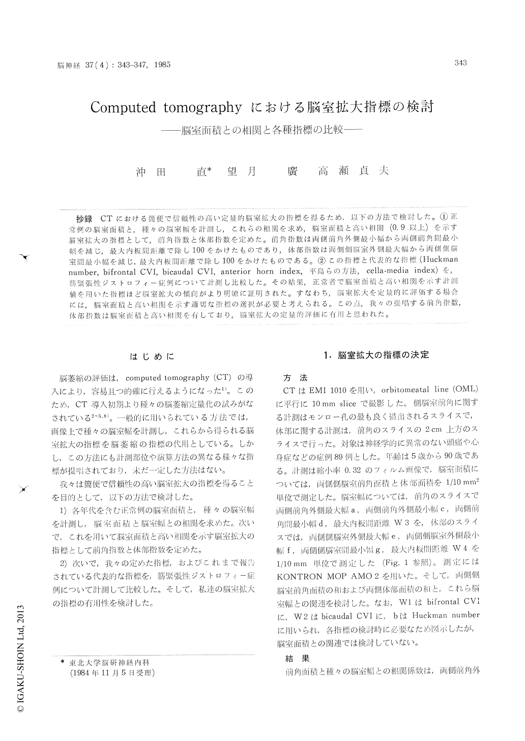
抄録 CTにおける簡便で信頼性の高い定量的脳室拡大の指標を得るため.以下の方法で検討した。①正常例の脳室面積と,種々の脳室幅を計測し,これらの相関を求め,脳室面積と高い相関(0.9以上)を示す脳室拡大の指標として,前角指数と体部指数を定めた。前角指数は両側前角外側最小幅から両側前角間最小幅を減じ,最大内板間距離で除し100をかけたものであり,体部指数は両側側脳室外側最大幅から両側側脳室問最小幅を減じ,最大内板間距離で除し100をかけたものである。②この指標と代表的な指標(Huckmannumber,bifrontal CVI,bicaudal CVI,anterior horn index,平島らの方法,cella-media index)を,筋緊張性ジストロフィー症例について計測し比較した。その結果,正常者で脳室面積と高い相関を示す計測値を用いた指標ほど脳室拡大の傾向がより明瞭に証明された。すなわち,脳室拡大を定量的に評価する場合には,脳室面積と高い相関を示す適切な指標の選択が必要と考えられる。この点,我々の提唱する前角指数,体部指数は脳室面積と高い相関を有しており,脳室拡大の定量的評価に有用と思われた。
CT scan is able to show cerebral atrophy more safely and more easily than pneumo-encephalo-graphy or cerebral angiography. Then, various methods have been reported for quantitative analysis of cerebral atrophy on CT scan. Gene-rally, cerebral atrophy might be judged from the ventricular dilatation with some indices, calculated from various ventricular width. But, there is no general agreement on what index is the most reliable. In this paper, we attempted to establish the index, easy to measure and most reliable. Our method is as follow.
Method
1) We carried out the CT scan (EMI 1010) on 89 neurologically intact patients. Scans were pa-rallel to orbito-meatal line (OML), and were 10 mm in thickness. On CT scan films, various width, area of anterior horns and area of bodies of late-ral ventricles were measured (Fig. 1). Measure-ment about the anterior horns of lateral ventri-cles were carried out on image the most clearly showed the foramen of Monro. And measurements about the bodies of lateral ventricles were on image, 20 mm above the image of anterior horn. Correlations of various width and areas were calculated (Table 1). Then we proposed new in-dices with high correlations (over O. 9) with vent-ricular area ; Anterior horn CVI (Cerebro-Vent-ricular Index) and Body CVI (Fig. 2, 3).
2) Patients with myotonic dystrophy show ce-rebral atrophy. We carried out the CT scan (GE-CT/T 8800) on 17 myotonic dystrophy patients and 30 controls. Between the two groups, age and sex were almost matched (Fig. 4). In the two groups, we calculated our new indices as well as various indices which have been reported ; Huck-man number, Bifrontal CVI, Bicaudal CVI, Ante-rior horn index, Hirajima's index, and Cella-me-dia index. The data were analyzed statistically (Table 2).
Results and Conclusion
The ventricular dilatation of myotonic dystrophy patients is more definite with Anterior horn CVI, Bicaudal CVI and Body CVI (p<0.01). These indices have higher correlations with the vent-ricular area (about 0.9).
From these results, we concluded that the re-liable index of ventricular dilatation on CT scan must have high correlation with the ventricular area, and our new indices (Anterior horn CVI and Body CVI) with high correlations with the ventricular area, are useful.

Copyright © 1985, Igaku-Shoin Ltd. All rights reserved.


