Japanese
English
- 有料閲覧
- Abstract 文献概要
- 1ページ目 Look Inside
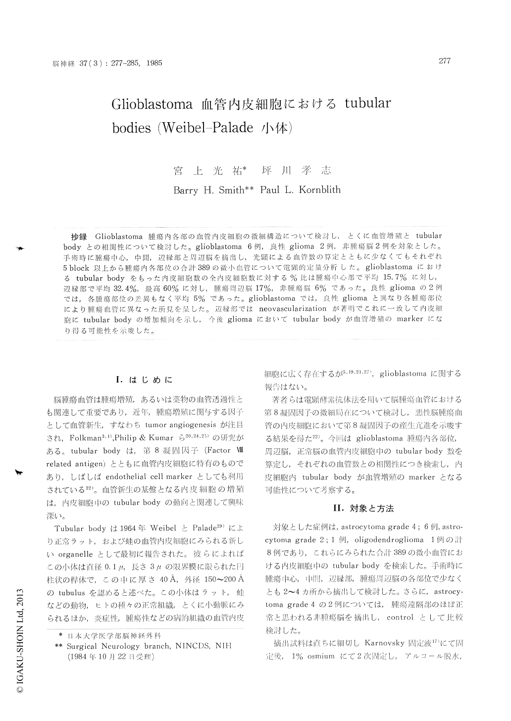
抄録 Glioblastoma腫瘍内各部の血管内皮細胞の微細構造について険討し,とくに血管増殖と tubular bodyとの相関性について検討した。glioblastoma 6例,良性glioma 2例,非腫瘍脳2例を対象とした。手術時に腫瘍中心,中間,辺縁部と周辺脳を摘出し,光顕による血管数の算定とともに少なくてもそれぞれ5 block以上から腫瘍内各部位の合計389の微小血管について電顕的定量分析した。glioblastomaにおけるtubular bodyをもった内皮細胞数の全内皮細胞数に対する%比は腫瘍中心部で平均15.7%に対し,辺縁部で平均32.4%,最高60%に対し,腫瘍周辺脳17%,非腫瘍脳6%であった。良性gliomaの2例では,客腫瘍部位の差異もなく平均5%であった。glioblastomaでは,良性gliomaと異なり各腫瘍部位により腫瘍血管に異なった所見を呈した。辺縁部ではneovascularizationが著明でこれに一致して内皮細胞にtubular bodyの増加傾向を示し,今後gliomaにおいてtubular bodyが血管増殖のmarkerになり得る可能性を示唆した。
Authors have studied the ultrastructure of endothelial cells in the microvessels of malignantand benign gliomas and in particular, the numbers of tubular bodies (Weibel-Palade) in endothelial cells of glioma microvessels in related with blood vessel proliferation. Glioblastoma 6, astrocytoma grade II 1, oligodendroglioma 1 and 2 samples of non-tumor brain tissue were analyzed quantitati-vely using light and electron microscope with Karnovski fixative.
All tissues were obtained from the center, the intermediate and the margin in each tumor tissue and justoutside of the tumor at operation. 389 microvessels were examined in the total glio-mas elctronmicroscopically.
Tubular body was first described by Weibel and Palade in the vascular endothelial cells of various organs in both man and animals. This is now con-sidered to be an organelle specific to the endothe-lial cell, but its function is still unknown. Tubular body observed in the endothelial cells of the glio-mas vessels consisted of a membrane-limited round, oval or elongated shaped intra cytoplasmic body (about 0.1-0. 2 rem) which contained tubules of 150-200 Å outer diameter. Tubular bodies were classified in the two types. One of them (mature type) was relatively electron dense to be more compact, the other (immature type) had relatively pale matrix. In the immature type they are loca-ted in close proximity to the Golgi complex or endoplasmic reticulum.
Mean % ratio of the endothelial cells including tubular bodies to whole endothelial cells were 15.7 %(center), 32.4% (margin), and 17% (justoutside) in the glioblastoma and 5% (non-tumor tissue). How-ever, mean % ratio of that in benign glioma were 5% and there were no difference in each localization.
Tubular bodies were common only in the endo-thelial cells in the marginal zones of glioblastoma with respect to the increased microvessels. In the endothelial cells of glioblastoma they are appeared consistent with neovascularity more prominently comparing with benign glioma. It is suggested that large numbers of tubular bodies may be a marker for proliferating endothelial cells.

Copyright © 1985, Igaku-Shoin Ltd. All rights reserved.


