Japanese
English
- 有料閲覧
- Abstract 文献概要
- 1ページ目 Look Inside
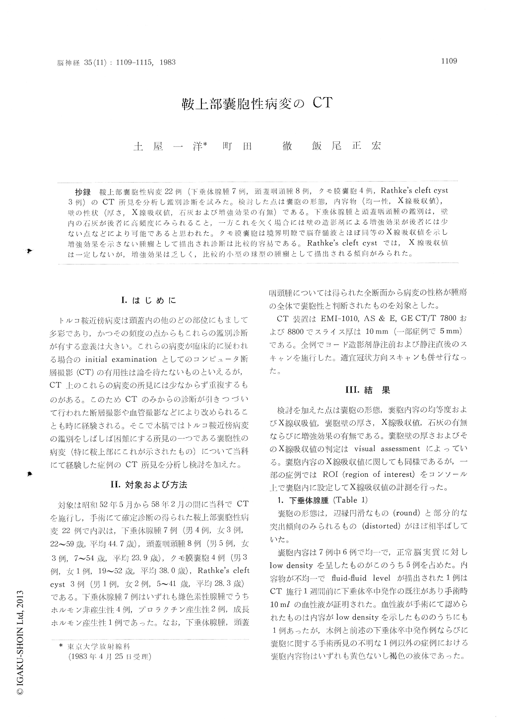
抄録 鞍上部嚢胞性病変22例(下垂体腺腫7例,頭蓋咽頭腫8例,クモ膜嚢胞4例, Rathke’s cleft cyst3例)のCT所見を分析し鑑別診断を試みた。検討した点は嚢胞の形態,内容物(均一性,X線吸収値),壁の性状(厚さ,X線吸収値,石灰および増強効果の有無)である。下垂体腺腫と頭蓋咽頭腫の鑑別は,壁内の石灰が後者に高頻度にみられること,一方これを欠く場含には壁の造影剤による増強効果が後者には少ない点などにより可能であると思われた。クモ膜嚢胞は境界明瞭で脳脊髄液とほぼ同等のX線吸収値を示し増強効果を示さない腫瘤として描出され診断は比較的容易である。Rathke's cleft cystでは,X線吸収値は一定しないが,増強効果は乏しく,比較的小型の球型の腫瘤として描出される傾向がみられた。
Difficulties are often encountered in the differen-tial diagnosis of suprasellar cystic lesions in computed tomography (CT). During the past 5 years and 7 months we experienced 22 such cases. In this report we tried to review the characteristic CT findings for the differential diagnosis of these lesions. Population of this study is consisted by 7 pituitary adenomas (4 non-functioning adenomas, 2 prolactinomas and 1 GH-secreting adenoma), 8 craniopharyngiomas, 4 arachnoid cysts and 3 Rathke's cleft cysts. In cases of pituitary adenomas and craniopharyngiomas, we included only those cases which were found to be completely cystic. Each case was scanned before and after contrast material injection and in majority of cases coronal scans were also obtained after contrast injection. The analysis was based on the CT appearance of the shape, the content and the wall of each cyst.The wall of the cyst was evaluated according to its thickness, density, presence of calcification and contrast enhancement. In 14 out of 22 cases the X-ray attenuation values of their content were calculated after setting the ROI in the cyst on the CT display console.
Craniopharyngioma often showed calcification in its wall, which was not seen in the wall of pitui-tary adenoma. The wall of pituitary adenoma revealed contrast enhancement in all cases, but half of craniopharyngioma showed no contrast enhancement in its wall. These two points are useful for the differential diagnosis of these lesions which we encounter most frequently. In addition, the mean X-ray attenuation value of the content of craniopharyngiomas (21. 3 H. U. in 3 cases) was lower than that of pituitary adenomas (27. 3 H. U. in 5 cases). The wall of 3 out of 7 cases of pitui-tary adenomas had locally distorted appearance but that of craniopharyngiomas seemed to be rounded. These findings may also help in the differential diagnosis of these lesions.
Arachnoid cysts are relatively easy to differen-tiate from the rest of suprasellar custic lesions. This is because the former were well delineated from the surrounding, showed almost equal X-ray attenuation value (9. 8 H. U. in 3 cases) to that of normal CSF, had round shape and showed no contrast enhancement. But in some cases of cranio-pharyngioma, especially if they lack calcification in the wall, and also in cases of low-density Rath-ke's cleft cyst, CT findings may be similar to those of arachnoid cyst.
The X-ray attenuation values of 3 cases of Rathke's cleft cyst varied (21. 4 H. U., 42. 7 H. U. and 64. 3 H. U., mean 42.8 H. U.). But they had tendency to be well-delineated and relatively small suprasellar masses with little contrast enhan-cement. These findings are suggestive of Rathke's cleft cyst.
In conclusion, CT findings of suprasellar cystic lesions in 22 cases were analyzed. Careful exami-nation of CT findings were found to be able to lead to the correct diagnosis of several suprasellar cystic lesions.

Copyright © 1983, Igaku-Shoin Ltd. All rights reserved.


