Japanese
English
- 有料閲覧
- Abstract 文献概要
- 1ページ目 Look Inside
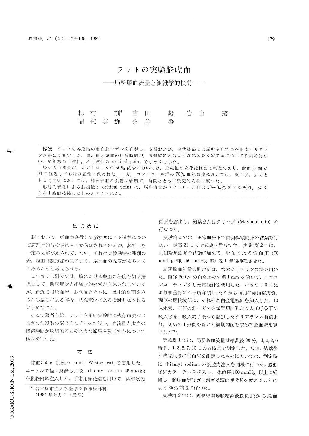
抄録 ラットの各段階の虚血脳モデルを作製し,皮質および,尼状核部での局所脳血流量を水素クリアランス法にて測定した。血流量と虚血の持続時間が,脳組織にどのような影響を及ぼすかについて検討を行ない,脳組織の可逆性,不可逆性のcritical pointを求めんとした。
局所脳血流量が,コントロールの50%減少においては,脳組織の変化は極めて軽微であり,虚血期間が21日経過してもほぼ正常に保たれた。一方,コントロール群の70%血流減少においては,虚血後,少くとも1時間後においては,神経細胞の損傷は著明で,時間とともに致死的変化に至つた。
It is supported that the neuronal damage resulted from cerebral ischemia depends on residual blood flow and or duration of ischemia. To determine whether the ischemic tissue would restore its fun-ction, the relationship among cerebral blood flow and histopathology in graded ischemia produced exprimentally, was investigated.
Varying degree of cerebral ischemia were pro-duced in adult Wistar rats by clipping of bilateral common carotid arteries either with or withoutinduced hypotention. rCBF was measured from bilateral caudate nuclei and parietal cortices using the hydrogen clearance method. Immediately after experiments, the brains were removed and fixed in formaline and stained by hematoxylin-eosin and the luxol fast blue methods to observe ischemic alternations.
In the control group, the mean value of rCBF was 46.6±4.9ml/100g/min (mean±SE) in the caudate nucleus and 48.3±4.6ml in the parietal cortex, respectively. The animals were divided into three experimental groups.
1) Immediately after clipping of bilateral com-mon carotid arteries, rCBF was reduced to an averrage of 25.4±1. 8 m/ in the caudate nucleus, and 25.4±2.1ml in the parietal cortex respectively, and was kept almost the same level through 21 experimental days. Histological abnormalities were absent at three hours after ischemic insult. Only dark staining of the cytoplasm in the cortex was present in a few experiments. In one or two day preparation, shrinkage and dark staining of the cytoplasm as well as microvacuolation around nerve cells were observed in some but not all expriments. However, these histological abnormalities disappear-ed in 21 days.
2) In rats which bilateral common carotid arteries were ligated and the blood pressure was reduced to 70 mmHg for four hous, rCBF was 12. 5±1. 7 ml in the parietal cortex and 14.1±1.0 ml in the caudate nucleus, respectively. In this group, ischemic cells were already detected in one hour and were particularly event in the entire cerebral cortex. Shrinkage as well as pyknosis were observed at three hours after ischemia and some nerve cells were surrounded by vacuolated neuropil.
3) In rats which bilateral common carotid arteries were ligated and the blood pressure was reduced to 50 mmHg for four hours, rCBF was not able to be measured in this method. Ischemic changes were more evident than that of the group 2. At two hours after ischemic insult, nerve cells in the cortex showed marked volumetric increase, blurring of the cytoplasmic boundaries, dark-blue staining of cytoplasm, and tortuous neurons. At four hours, pyknosis of nerve cells were observed in a large proportion of the neurons.
From these results, it is suggested that the brain tissue tolerates 50per cent reduction of rCBF and is able to restore its function after ischemic insult. On the contrary, seventy per cent reduction of rCBF produced severely injured brain even in the early stage of ischemia.

Copyright © 1982, Igaku-Shoin Ltd. All rights reserved.


