Japanese
English
- 有料閲覧
- Abstract 文献概要
- 1ページ目 Look Inside
1.はじめに
脳神経外科領域においてトルコ鞍に異常をきたす疾患は数多くあり,トルコ鞍拡大はそれのみで下垂体腫瘍あるいはトルコ鞍近傍病変を示す重要な所見である1,2,6,10,14,19,23〜25)。一方,近年内分泌診断学の進歩により下垂体マイクロアデノーマへの関心が一層深まつている。その診断基準としては鞍底の変化は別として,トルコ鞍の容積が正常範囲内であることが条件とされている15)。したがつてトルコ鞍容積の正常範囲を明確にするため,種々のトルコ鞍計測値の正常上限値が必要とされる。トルコ鞍容積についての報告は19世紀末のHrdlika24)の算出法に端を発し,20世紀前半のBalli,Bussi,Haas,Sche—uermann30),Pandoroso24)らに続き,Dichiro6,7),Taveras29)らの計測法がよく知られている。以上のごとく欧米においては,トルコ鞍計測上の正常域はほぼ確立されているが,本邦においては過去に2,3の試み18,28)はあるが多数例を詳細に検討した報告はまだ見られない。しかも,長頭型の欧米人の基準を短頭型の日本人にそのまま適用することは必ずしも妥当ではないと考えられる。今回われわれは,正常頭蓋単純撮影1,000例について,トルコ鞍の計測および形態分類とそれらについての統計学的検討を行つたので報告する。
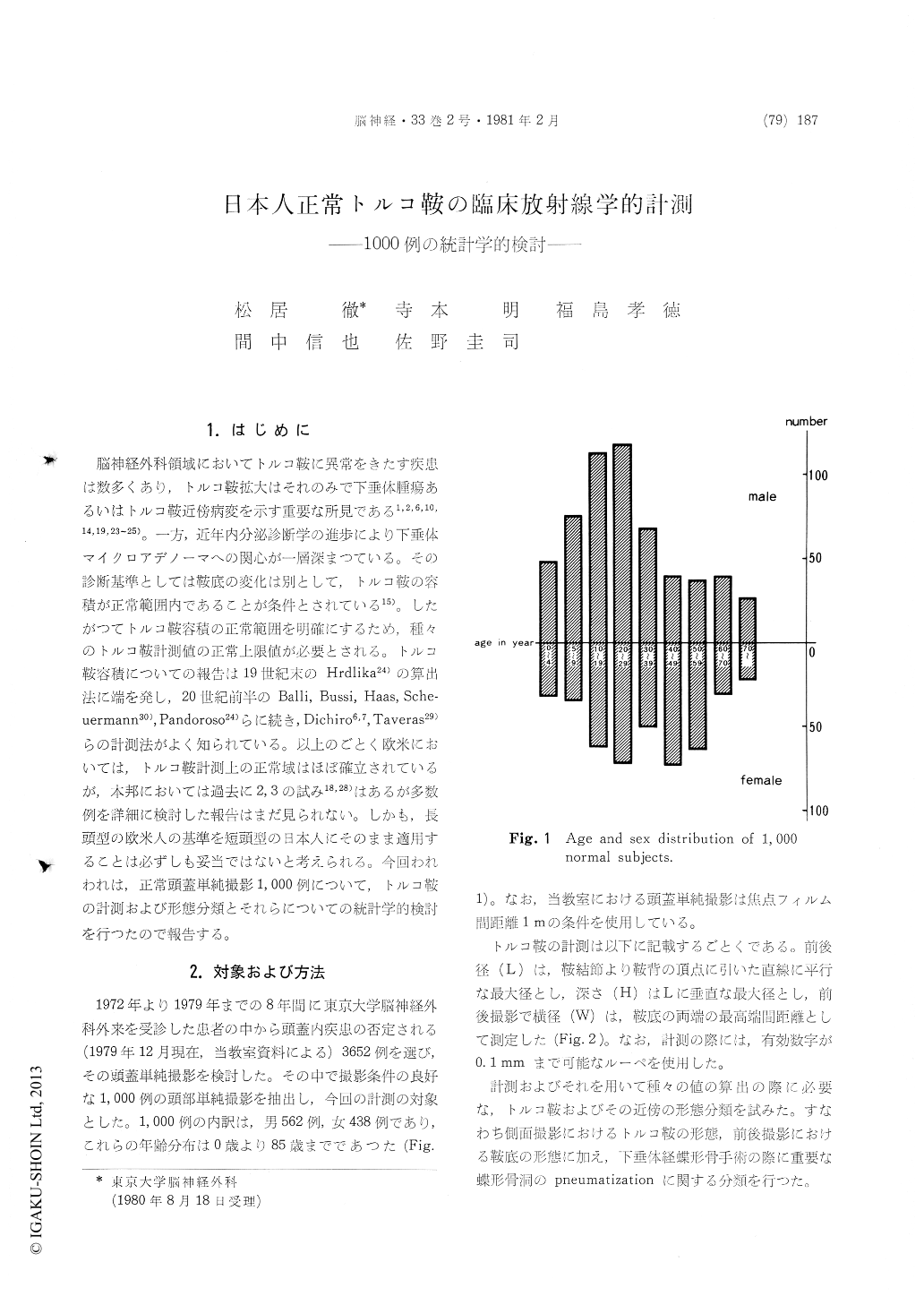
Enlargement of the sella turcica on the X-ray films is important finding which by itself suggests some sellar and parasellar pathology. On the other hand, it is the essential condition for the diagnosis of pituitary "microadenomas" that the sella turcica is of normal size without ballooning. Thus, it becomes the crucial point to know the normal range of the sellar size. The normal range of the di-mensions of the sella turcica among caucasians has been well known through the works of DiChiro or of others, Among Japanese people, however, the normal upper limit of the sellar size on radiogra-phical measurements have not yet been established. The present work has been undertaken to define the normal values of the sellar size in the Japanese using the skull X-ray films of 1,000 subjects who have no known neurological diseases. The age of the subjects ranged from 0 to 85, 562 of them being males and the rest females. The A-P length (L), the height (H) and the width (W) of the sella turcica were measured in lateral and postero-anterior projections. The sellar valume and square were calculated through our formula using above three measurements. In addition, the forms of sella turcica and sphenoid sinus in lateral projection and configuration of the sellar floor in postero-anterior projection were classified into several types. Our results can be summerized as follows;
1) The configuration of the sellar floor in postero-anterior projection were classified into 6 types.
1) flat: 54.9%
2) inclined: 14.3%
3) concave: 11.2%
4) convex: 9.6%
5) V-shaped: 9.0%
6) reversed V-shaped: 0.3%
2) The forms of the sella turcica in lateral pro- jection were classified into 5 categories.
1) U-shaped: 64.3%
2) oval: 21.2%
3) global: 9.0%
4) flat: 5.0%
5) J-shaped: 0.6%
3) The degree of pneumatization of the sphenoid sinus was divided into 3 types. The airation of sphenoid sinus was found to become promi- nent with increasing age.
1) concha: 3.2%
2) presellar: 17.0%
3) sellar: 79.8%
4) Some expansive apperance of the sella turcica like ballooning was noted in 3.3% of the normals and erosive changes of the sellar floor in 3.5%. The double floor was found in 7.2%.
5) The normal range of the three dimensional values of the sella turcica in adults were
L: 12.0±3.5mm (mean±2SD)
H: 8.9±2.6mm
W: 14.5±5.8mm
The calculated volume and square of the sella turcica using our formula were highly correlated with the above values, so that the size of the sella can be simply represented by L or H. The normal upper limits of 'L' and 'H' are 15.5 mm and 11.5mm respectively.
Copyright © 1981, Igaku-Shoin Ltd. All rights reserved.