Japanese
English
- 有料閲覧
- Abstract 文献概要
- 1ページ目 Look Inside
はじめに
眼静脈・海綿静脈洞撮影は1951年Dejeanが報告して以来,種々の方法をもつて行なわれるようになり,眼窩内及びトルコ鞍近傍病変の診断に対してその有用性が論じられている。著者は本撮影を1969年11月より46症例に対して,主として前頭静脈の経皮的穿刺法により行なつてきた。今回著者の行なつている造影法を述べるとともに,主としてトルコ鞍近傍病変に対する本撮影の診断的価値を,海綿静脈洞の変形ならびに計測により検討した。その結果本撮影の有用性を認めたので報告する。
In the neuroradiological diagnosis of the sellar or parasellar tumors, carotid angiography and pneumoencephalography have been widely applied. By these methods, detection of the suprasellar ex-tension of these tumors is possible, but detection of the intrasellar or lateral extension of the tumors is very difficult.
For more precise diagnosis of these tumors the cavernous sinus venography was performed in 46 cases over the past two years. Eighteen cases of them were pituitary tumors, 11 were parasellar tumors and two were orbital tumors. There were twelve cases in which no abnormalities were de-tected in the cellar or parasellar regions by veno-graphic study.
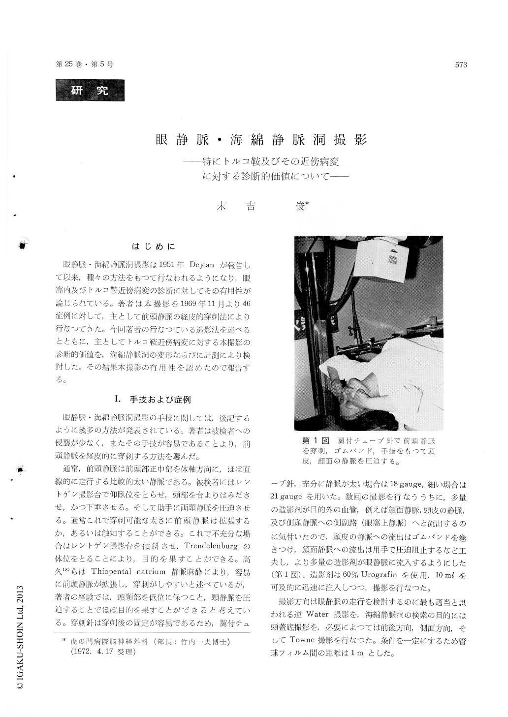
Under head down position, a 18 to 20 gaugeneedle was percutaneously introduced into the frontal vein and 8 to 10 ml of 60% Urografin was injected as fast as possible. Then X-ray pictures of the basel and reverse Water's view were taken immediately after injection.
In this paper the diagnostic value of this method in regard to the sellar or paraseller tumors was mainly discussed. Definite abnormalities such as venous occlusion, displacement and deformity of the cavernous sinus were observed. The minimumintercavernous distance between both cavernous sinuses was measured. The minimum intercavern-ous distance in normal eleven cases was 12 mm to 20 mm (average 16 mm). In all cases of the pitui-tary tumor it was over 30 mm (average 37 mm). The result of this study shows that the cavernous sinus venography is the most valuable examination for detection of the state of the lateral extension of the sellar and parasellar tumors.

Copyright © 1973, Igaku-Shoin Ltd. All rights reserved.


