Japanese
English
- 有料閲覧
- Abstract 文献概要
- 1ページ目 Look Inside
I.はじめに
Gargoylismはdwarfism, deafness, splenohepato—megaly,四肢屈曲等を主徴とし,その本態としてmuco—polysaccharidosisとgangliosidosesの両面を兼ね備えた疾患である。本疾患に水頭症が合併することは,よく知られているが,最近我々は,このような症例に対しV-A shuntを行なう機会を得たので若干の考察を加えて報告する。
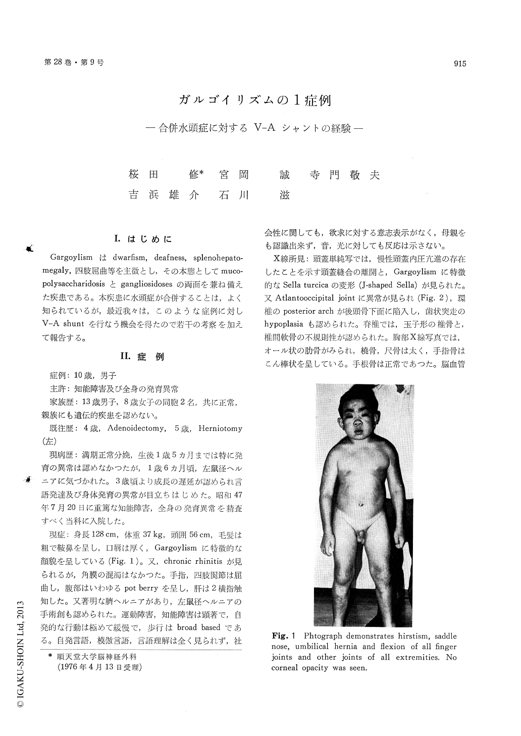
A ten-year-old boy was first examined at our Department in July, 1972 because of his mental retardation and generalized somatic anomaly. The boy was not able to talk and understand simple conversation and showed no interest in his envi-ronment. Hirsutism, saddle nose, umbilical hernia and flexion of all finger joints and other joints of all extremities were noted. No corneal opacity was seen. The liver edge was palpable two finger breadth below the costal margin. X-ray studies of the skull showed separation of the cranial sutures, J-shaped sella, hypoplasia of odontoid and intrusion of posterior arches of atlas into posterior aspect of the foramen magnum. The PEG showed markedly dilated ventricle and poor filling of subarachnoid space. Excessive acid mucopolysaccharide was ex-creted in the urine.
On August 21, 1972, V-A shunt was carried out. Difficulties were met during intubation because of the anomaly of his oral cavity and NLA was carried out instead. Ventricular pressure at the time of surgery was 550 mmH2O. Right after the surgery tracheotomy was carried out because of the post-operative development of pneumonia. After im-provement of general condition and extubation, he spoke a few monosyllabic words. On September 9, 1972, he suddenly developed dyspnea and tracheo-tomy was performed again. Thereafter, bilateral hyperreflexia gradually developed. GAG revealed bilateral subdural hematoma, and these were irrigat-ed. He suddenly died on June 28, 1973, with dyspna.
It should be stressed that in the patients of Gargoylism a simple surgical procedure like V-A shunt or even more anesthesia are followed by the various complications.
Ultrastructures of the cerebral biopsy in this case were characterized as follow : 1) granulomembranous body 2) zebra body 3) structure similar to membra-nous cytoplasmic body.

Copyright © 1976, Igaku-Shoin Ltd. All rights reserved.


