Japanese
English
- 有料閲覧
- Abstract 文献概要
- 1ページ目 Look Inside
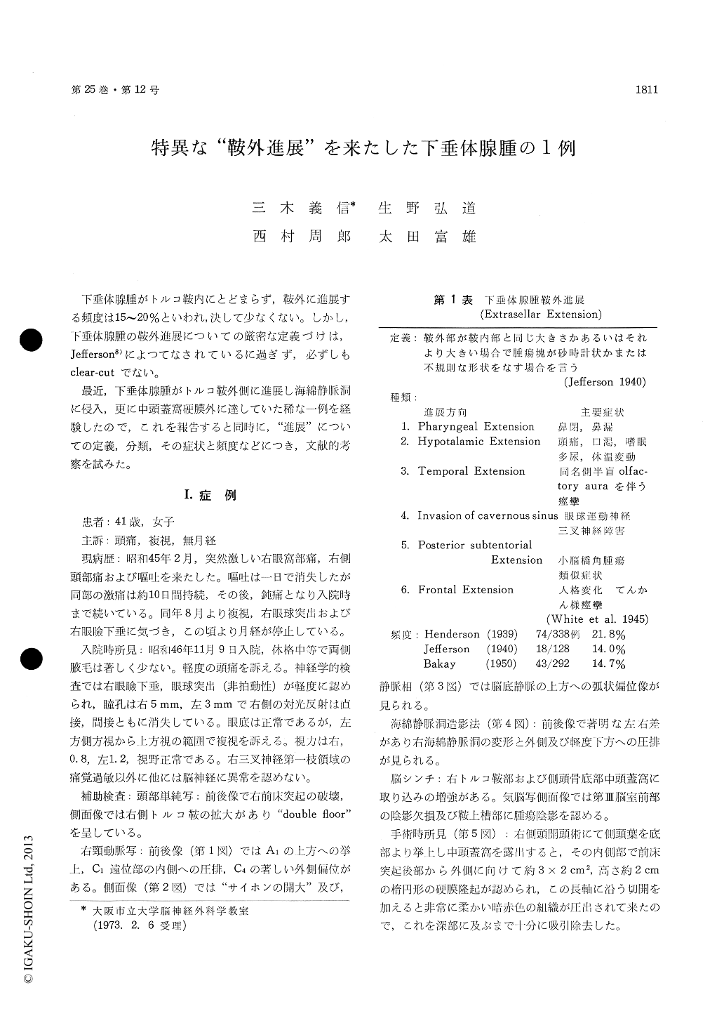
下垂体腺腫がトルコ鞍内にとどまらず,鞍外に進展する頻度は15〜20%といわれ,決して少なくない。しかし,下垂体腺腫の鞍外進展についての厳密な定義づけは,Jefferson8)によつてなされているに過ぎず,必ずしもclear-cutでない。
最近,下垂体腺腫がトルコ鞍外側に進展し海綿静脈洞に侵入,更に中頭蓋窩硬膜外に達していた稀な一例を経験したので,これを報告すると同時に,"進展"についての定義,分類その症状と頻度などにつき,文献的考察を試みた。
Forty one-year-old house wife felt severe pain suddenly in the right orbital and temporal region and vomiting has appeared at the same time tenmonths prior to admission. Although vomiting has disappeared on the next day, headache has been persistent about ten days thereafter. Sub-sequently she has been improving but dull pain in the right frontal region has been present until her admission. Six months after the onset of the symptom, diplopia, eyelid ptosis and exophthalmos has appeared, then her menses has stopped.
Findings on admission were as follows : Scant axillary hair and amenorrhea. Eyelid ptosis and slight exophalmos on the right side. Pupil was 5mm on the right side and 3 mm on the left side. No light reflex was noted on the right side and visual acuity was 0.8 on the right and 1.2 on the left. Ocular fundi and visual field were normal. Diplopia was noted on upward and left lateral gaze. Hyperalgesia was present in the area in-nervated by the opthalmic nerve on the right side. Plain craniogram showed destruction of the anterior clinoid process and "double floor" due to unilateral enlargement of the sella turcica. CAG : A-P viewrevealed upward displacement of the A-1, inward shift of the distal portion of the C-1 and outward shift of the C-4. The lateral arteriogram showed opening of the syphon and archlike upward dis-placement of the basilar vein of Rosenthal was noted on the lateral phlebogram. Deformation and lateralward shift of the right cavernous sinus was noted on the cavernous sinogram. The temporal craniotomy revealed a greyish pink soft tumor of walnut size originating in the sella turcica and extending into the right middle fossa between the dura mater and the basal bone. The tumor was extirpated by suction through the dural incision made over the mass. Histological diagnosis was chromophobe adenoma of the pituitary gland. Her menses has appeared three months after the surgery.
Definition, classification, incidence and sympto-matology of the extrasellar extension of a pituitary adenoma as well as its pertinent diagnostic pro-cedures were discussed.

Copyright © 1973, Igaku-Shoin Ltd. All rights reserved.


