Japanese
English
- 有料閲覧
- Abstract 文献概要
- 1ページ目 Look Inside
I.はじめに
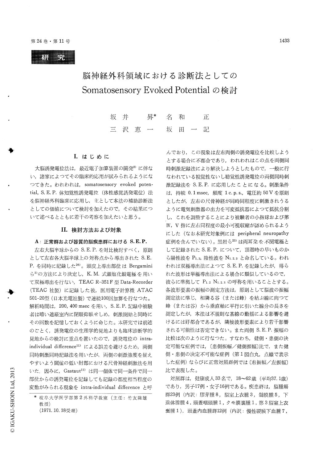
大脳誘発電位法は,最近電子加算装置の開発8)に伴ない,諸家によつてその臨床的応用が試みられるようになつてきた。われわれは,somatosensory evoked poten—tial, S.E.P.体知覚性誘発電位(体性感覚誘発電位)法を脳神経外科臨床に応用し,主として本法の補助診断法としての価値について検討を加えたので,その結果について述べるとともに若干の考察を加えたいと思う。
Clinical application of somatosensory evoked potential (SEP) examination in neurosurgical cases was studied. Bilateral ulnar nerves were simulta-neously stimulated and bilateral SEPs were recorded bipolarly from the scalp surfaces corresponding to somatosensory areas. Peak latency and ratio of amplitude between both sides were measured on various components of SEP in 33 normal subjects and in 45 cases of various organic brain diseases. Results obtained were as follows.
1) Forty-five cases of various organic brain dis-eases were classified into tumor (T), hematoma (H) and cerebrovascular lesion (CVL) groups. Peak latency tended to be prolonged in all 3 groups, as compared with normal control. This tendency was marked on the pathological side and, in T-group, it was especially marked in late components. Ratio of amplitude on the lesion side to that on the healthy side showed the following tendency. It was often larger than 1.0 in N1 and smaller than 1.0 in N2 in T-group. In the other 2 groups, it was smaller than 1.0 in both Ni and N2, except that in some cases of chronic subdural hematoma the ratio of amplitude in N2 was almost equal to or larger than 1.0.
2) T-group was divided into malignant and benign subgroups on the histological ground. Tendency of peak latency delay and tendency of higher amplitude of N1 on the lesion side were more marked in the malignant subgroup.
3) From the point of view of tumor localization, SEP changes were most marked in the cases of parieto-temporal or thalamic tumors and no SEP change was observed in the cases of tumors in the chiasmal region.
4) In the cases of unilateral sensory disturbance of central origin, peak latencies of N1 and N2 tended to be prolonged and reduction in amplitudes of N1and N2 (especially N1) on the lesion side was cha-racteristic, except that in a few tumor cases N1 was of higher amplitude on the lesion side despite presence of sensory disturbance.
5) In the course of recovering sensory disturbance, it was generally observed that the amplitudes of N1 and N2 on the lesion side, which had been for-merly reduced, increased to regain normal SEP pattern. This phenomenon was most typical in the cases of intracranial hematoma, especially of chronic subdural hematoma. In the tumor cases, in which N1 amplitude was higher on the lesion side despite presence of sensory disturbance, it was observed that N1 amplitude was quickly normalized following tumor extirpation in parallel with clinical improve-ment.
6) Finding of EEG slowing tended to show a parallel relationship with peak latency delay and amplitude reduction on the lesion side in SEP. In not a few cases with normal EEG, abnormal SEP findings were observed.
Studies on recovery function of human somato-sensory evoked potential were performed in 10 normal control subjects and in 30 cases of posttr-aumatic chronic headache. Relationship between these results and scores of Cornell Medical Index (C. M. I. according to Fukamachi modification) was examined. Results obtained were as follows.
7) In control subjects, amplitude of SEP to test stimulus increased gradually as the interval between conditioning and test stimuli was increased from 50 to 300 msec. Approximate 100% recovery was attained at an inter-stimulus interval of 200 msec as for N1 and at 300msec as for N2 and P2.
8) Recovery function curve of N2 in posttraumatic headache cases showed any one of the following 3 types i) supernormal recovery at around 100 msec, ii) supernormal recovery at around 200 msec and iii) lower recovery function as a whole than the control. In most of the posttraumatic headache cases classified as area I and II of C. M. I., it was seen that facilitation was remarkably present when the test stimulus was given 100 or 200 msec after the conditioning one. In most of the cases classified as area IV of C. M. I. no facilitation was present and values of recovery function generally were lower than the normal control. In the cases of area III of C. M. I., facilitation was or was not present. The above findings were observed also in P2. Effects of stellate ganglion blockade on recovery function of SEP were examined. Recovery function at an inter-stimulus interval of 100 msec in the cases of area I and II of C. M. I. was compared with that in the cases of area IV of C. M. I., the latter serving as the control, and the following resultswere obtained.
9) In the control group, stellate ganglion blockade generally caused an increase in amplitude of N2. The cases of area I and II of C. M. I., in which stellate ganglion blockade was effective clinically, also showed similar tendency.
10) In the control group, in which the blockade was usually ineffective clinically, recovery function of N1, N2 and P2 was not affected significantly bythe blockade, while in the cases of area I and II in which the blockade was effective clinically, the phenomenon of supernormal recovery observed originally at an inter-stimulus interval of 100msec disappeared and the recovery function was obser-ved to be normalized after the blockade.
These results obtained were discussed.

Copyright © 1972, Igaku-Shoin Ltd. All rights reserved.


