Japanese
English
- 有料閲覧
- Abstract 文献概要
- 1ページ目 Look Inside
はじめに
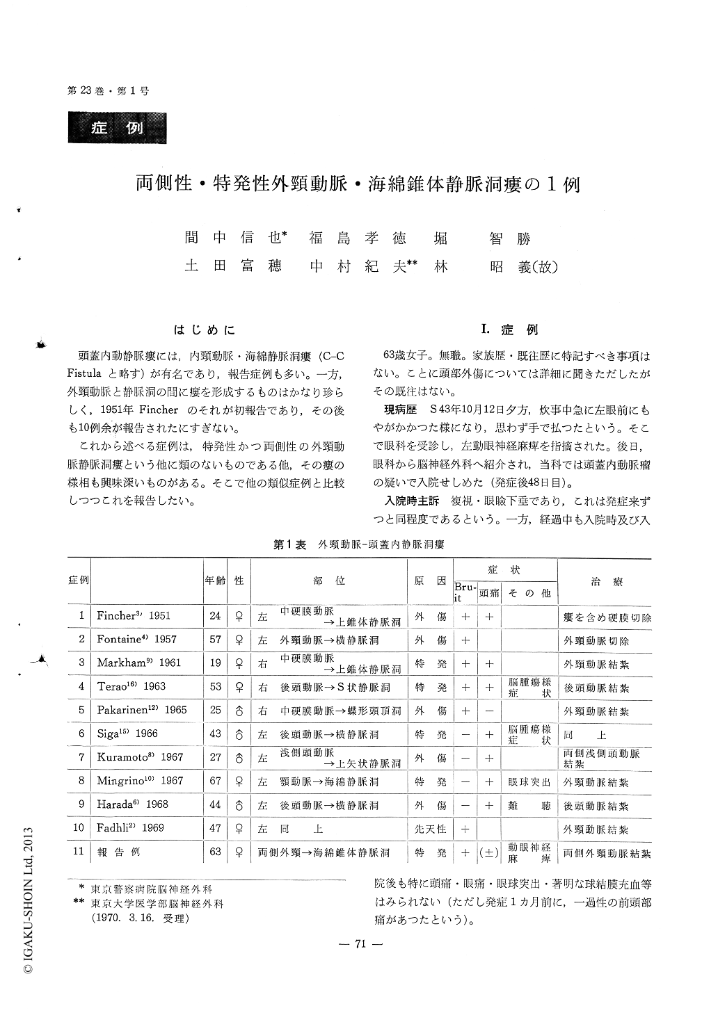
頭蓋内動静脈瘻には,内頸動脈・海綿静脈洞瘻(C-CFistulaと略す)が有名であり,報告症例も多い。一方,外頸動脈と静脈洞の間に瘻を形成するものはかなり珍らしく,1951年Fincherのそれが初報告であり,その後も10例余が報告されたにすぎない。
これから述べる症例は,特発性かつ両側性の外頸動脈静脈洞瘻という他に類のないものである他,その瘻の様相も興味深いものがある。そこで他の類似症例と比較しつつこれを報告したい。
An obese, slightly hypertensive 63-year-old woman was healthy, untill abrupt manifestation of left oculomoter palsy. No history of head injury wasremenbered. The palsy was the only symptome at the begining. However, 62 days later, she noticed intracranial murmur. Headache, exophthalmos con-junctival injection and papilledema were all negative in the clinical course.
On admission, common carotid angiogram appeared just like C-C fistula, but selective external carotid angiogram on both side revealed carotid-caverno-petrosal, pistula supplied from right as well as left external carotid arteries simultaneously.
After the first trial of clipping of the left meningel artery, temporaly aggravation of the oculomotorpalsy was observed. We postulated that unfavour-able charge of hemodynamic influence directly to the intracauernous portion of the oculomotor nerve resulted in such an aggravation.
We treated the patient in success finally by bilateral ligation of external carotids at the neck. At discharge, intracranial murmur as well as left oculomoter palsy disappeared except for slight my-driasis. One-year follow-up demonstrates no further recurrence.

Copyright © 1971, Igaku-Shoin Ltd. All rights reserved.


