Japanese
English
- 有料閲覧
- Abstract 文献概要
- 1ページ目 Look Inside
I.はじめに
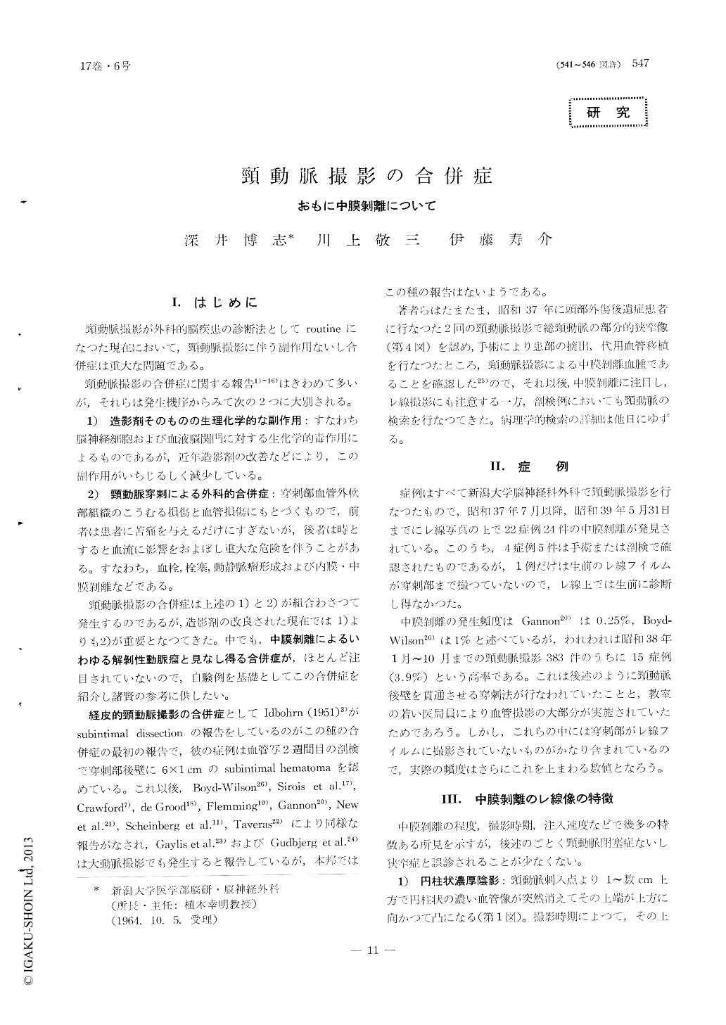
頸動脈撮影が外科的脳疾患の診断法としてroutineになつた現在において,頸動脈撮影に伴う副作用ないし合併症は重大な問題である。
頸動脈撮影の合併症に関する報告1)〜16)はきわめて多いが,それらは発生機序からみて次の2つに大別される。
As a complication of carotid angiography, mechanical injury of the arterial wall is important. Recently, we have experienced the medial dissection and/or valve formation of the arterial wall in 24 instances with 22 patients during the last two years. The incidence was about 4% of carotid angiography performed. A few attentions have been payed to such complications pre-viously, because of its rare clinical manifestations. Our studies show, however, such complications are not so infrequent as previously noted.
X-ray films of the case with the dissection may reveal several characteristic features, which were classified into 5 types. After the histological examination on 5 lesions of 4 cases, it was noted that such arterial wall dis-section always occured in the medial muscular layer alone. Hematoma formation within in the muscular layers, valve formation and dissecting aneursm were their major pathological evidences.
The most possible mechanism of such undesirable process could be as follows:when the bevelled lumen of the needle is partly located in the carotid lumen and partly located within the posterior wall of the caroitd artery, pulsatile backflow must be occured through the needle. Under such condition, the forceful injection can produce more or less an intramural extravasation of contrast medium, with extensive arterial wall dissection.
Fortunately, no detectable clinical symptoms were encountered in our series. However, it should be always emphasized that the medial disssection may result in serious complications in the aged subjects or patients with the atheromatous carotid artery. As the prevention of such serious complications, it was con-cluded, therefore, that the most importamt point is to avoid the penetration (damage) of the posterior wall of the carotid artery.

Copyright © 1965, Igaku-Shoin Ltd. All rights reserved.


