Japanese
English
- 有料閲覧
- Abstract 文献概要
- 1ページ目 Look Inside
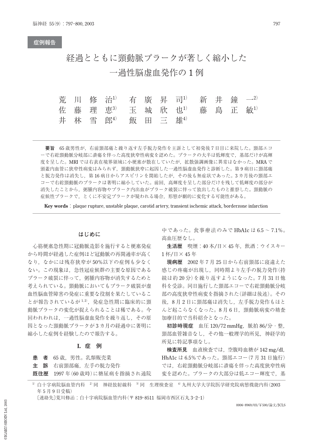
要旨 65歳男性が,右前頸部痛と繰り返す左手脱力発作を主訴として初発後7日目に来院した。頸部エコーで右総頸動脈分岐部に潰瘍を伴った高度狭窄性病変を認めた。プラークの大半は低輝度で,基部だけが高輝度を呈した。MRIでは右表在境界領域に小梗塞が散在していたが,拡散強調画像に異常はなかった。MRAで頭蓋内血管に狭窄性病変はみられず,頸動脈狭窄に起因した一過性脳虚血発作と診断した。第9病日に頸部痛と脱力発作は消失し,第16病日からアスピリンを開始したが,その後も無症状であった。3カ月後の頸部エコーで右総頸動脈のプラークは著明に縮小していた。前回,高輝度を呈した部分だけを残して低輝度の部分が消失したことから,粥腫内容物やプラーク内出血がプラーク破裂に伴って放出したものと推察した。頸動脈の症候性プラークで,とくに不安定プラークが疑われる場合,形態が劇的に変化する可能性がある。
Abstract
A 65-year-old man visited to our hospital, because he felt dull pain on the right side of the neck and had transient weakness of the left hand repeatedly. Carotid ultrasonography revealed a large plaque with severe stenosis and ulceration at the bifurcation of right common carotid artery. The plaque was mostly hypoechoic and only the basal part was hyperechoic. Magnetic resonance (MR) study showed multiple high-intensity spots in superficial borderzone area of the right cerebral hemisphere on fluid attenuated inversion recovery images, although diffusion-weighted images revealed no abnormality. Intracranial stenotic lesion was not detected by MR angiography. We diagnosed transient ischemic attacks due to right carotid artery stenosis. Nine days after the symptom onset, he showed no signs of cerebral ischemic attacks and seven days later he began taking aspirin and remained stable. Three months later, carotid ultrasonography showed marked shrinkage of the carotid plaque, of which hypoechoic part virtually disappeared. Release of the atheroma gruel and/or intraplaque hemorrhage due to plaque rupture might lead to dramatic change of the carotid plaque.

Copyright © 2003, Igaku-Shoin Ltd. All rights reserved.


