Japanese
English
- 有料閲覧
- Abstract 文献概要
- 1ページ目 Look Inside
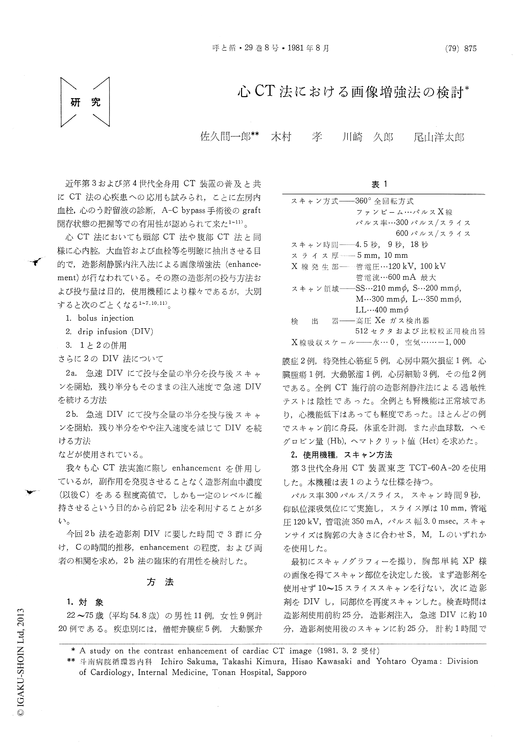
近年第3および第4世代全身用CT装置の普及と共にCT法の心疾患への応用も試みられ,ことに左房内血栓,心のう貯留液の診断,A-C bypass手術後のgraft開存状態の把握等での有用性が認められて来た1〜11)。
心CT法においても頭部CT法や腹部CT法と同様に心内腔,大血管および血栓等を明瞭に抽出させる目的で,造影剤静脈内注入法による画像増強法(enhance—ment)が行なわれている。その際の造影剤の投与方法および投与量は目的,使用機種により様々であるが,大別すると次のごとくなる1〜7,10,11)。
Computed tomography (CT) is considered to be applicable to heart and its usefulness has been reported particulary in recognition of interatrial thrombi and pericardial effusion. For detailed examination of heart by CT, it is necessary to enhance the contrast of CT image by intravenous administration of contrast medium.
Presently, both of plain and contrast enhanced CTs were carried out on 20 patients with cardiac disease. For contrast enhancement of the CT images, 220 ml of 30% meglumine iotalamate (30% DIP CONRAY) was administered by drip intravenous infusion (DIV) : 110 ml as an initial loading dose and 110 ml as a subsequent mainte-nance dose. In this study, 3 different protocols for the timing of DIV were examined : A ; 3. 5to 5 and 9 to 15 minutes, B ; 5. 5 to 7 and 15 to 25 minutes, and C ; 2. 5 and 15 minutes, for load-ing and maintenance respectively. Plasma con-centration of meglumine iotalamate (C) was determined by UV method and its temporal changes during DIV were investigated. The degree of contrast enhancement was evaluated by the difference of CT numbers within the heart between before and during DIV.

Copyright © 1981, Igaku-Shoin Ltd. All rights reserved.


