Japanese
English
- 有料閲覧
- Abstract 文献概要
- 1ページ目 Look Inside
- 参考文献 Reference
- サイト内被引用 Cited by
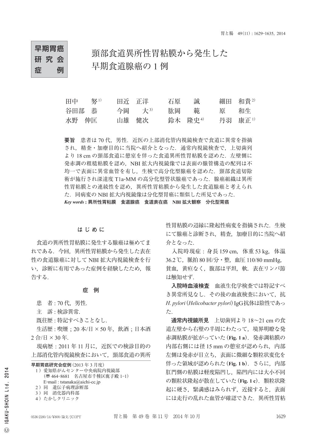
要旨 患者は70代,男性.近医の上部消化管内視鏡検査で食道に異常を指摘され,精査・加療目的に当院へ紹介となった.通常内視鏡検査で,上切歯列より18cmの頸部食道に憩室を伴った食道異所性胃粘膜を認めた.左壁側に発赤調の粗糙粘膜を認め,NBI拡大内視鏡像では表面の腺管構造の配列は不均一で表面に異常血管を有し,生検で高分化型腺癌を認めた.頸部食道切除術が施行され深達度T1a-MMの高分化型管状腺癌であった.腺癌組織は異所性胃粘膜との連続性を認め,異所性胃粘膜から発生した食道腺癌と考えられた.同病変のNBI拡大内視鏡像は分化型胃癌に類似した所見であった.
A 70-year-old man was referred to our hospital for further examination of an esophageal lesion that had been incidentally identified by EGD(esophagogastroduodenoscopy). EGD revealed a reddish and coarse mucosa in the left side of the heterotopic gastric mucosa of the cervical esophagus approximately 18cm from the teeth. ME-NBI(magnifying endoscopy with narrow band imaging)showed irregular microsurface structures and vessels. The coarse mucosa was diagnosed as well-differentiated adenocarcinoma by endoscopic biopsy. Resection of the cervical esophagus was performed. The pathological diagnosis was well-differentiated adenocarcinoma, and the tumor was located in the muscularis mucosae. Histological analysis identified the esophageal adenocarcinoma as arising from the heterotopic gastric mucosa. The ME-NBI appearance of this tumor resembled that of a differentiated gastric cancer.

Copyright © 2014, Igaku-Shoin Ltd. All rights reserved.


