Japanese
English
- 有料閲覧
- Abstract 文献概要
- 1ページ目 Look Inside
胃の平滑筋肉腫は悪性リンパ腫を含む胃の非上皮性悪性腫瘍の約20%を占めるとされており1),今日それほど稀な疾患ではないが,術前診断率はまだ低い現状にある1)2).術前診断が困難なのは他の疾患と鑑別できる特徴的な所見が少ないためといわれている7).今回,われわれは特異な胃X線像,ならびに腹部血管造影像より術前に胃平滑筋肉腫と想定しえた1例を経験したので報告する.
A report is made of gastric leiomyosarcoma diagnosed preoperatively by characteristic radiologic features of the stomach.
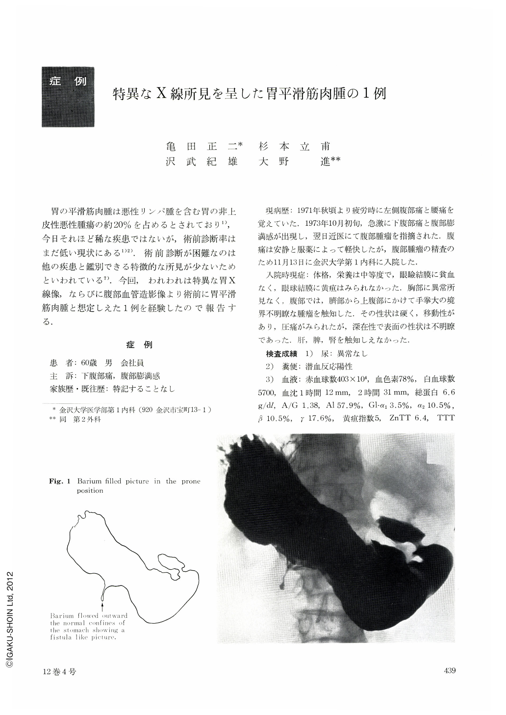
A 60 years old man was referred to our hospital because of lower abdominal pain and fullness. Physical examinations on admission revealed a tumor the size of a man's fist in the upper part of the abdomen. It was firm, tender, and movable. Laboratory findings were abnormal only in that stool specimen gave positive reaction with hematest. X-ray examination of the stomach showed extrinsic pressure on the greater curvature of the corpus. From the central point of the compressed area, barium flowed outward from the normal confines of the stomach, showing a fistulalike picture. The mucosal pattern appeared normal. Endoscopic studies revealed a fistula-like picture toward which mucosal folds converged. Endoscopic retrograde pancreaticograph disclosed no abnormality in the pancreas duct. Celiac arteriography disclosed a richly vasculized stellate tumor supplied by left gastric artery. Under a diagnosis of probable gastric leiomyosarcoma, laparotomy was performed. A soft exogastric tumor, 17×13×13 cm, was identified on the greater curvature of the corpus. There was a pseudo-diverticulum in the stomach wall which led into the tumor mass. Histologic specimens established a diagnosis of gastric leiomyosarcoma.
Most authors in the past emphasized that accurate preoperative diagnosis of gastric leiomyosarcoma is difficult because of no pathognomic symptoms and signs, laboratory criteria, or radiologic features. However, from our case described above, we consider it possible to make preoperative diagnosis in certain groups of gastric leiomyosarcoma by characteristic radiologic features.

Copyright © 1977, Igaku-Shoin Ltd. All rights reserved.


