Japanese
English
- 有料閲覧
- Abstract 文献概要
- 1ページ目 Look Inside
癌はほとんど全胃に拡がっていながら胃集検のレベルでは恐らく発見困難であったろうと思われる平坦型早期胃癌で,かつその癌巣の拡がりが,X線検査,内視鏡検査の所見および切除標本の肉眼所見でも判然としなかった症例を報告する.
A case of flat type early gastric cancer is described that would surely have escaped detection on a routine level of mass screening. The horizontal extent of cancer spread defied not only X-ray and endoscopy but gross observations of the resected specimens as well.
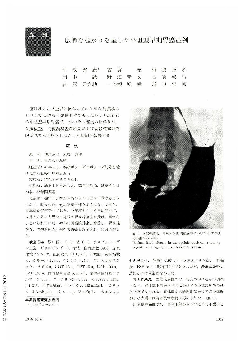
A man aged 54 had undergone gastric mass screening every year with impunity, but heavy feeling after meals led him to us for further check-up of the stomach. Initial X-ray revealed marginal rigidity and irregularity from the angle down to the pyloric antrum. Further detailed X-ray in barium-filled picture disclosed likewise rigid and irregular contour along both curvatures from the level of the angle to the pyloric antrum. Double contrast study revealed indistinct gastric areae and radiolucent round shadows suggesting nodular protrusions. These findings were scattered about over an extensive area, but we were unable to make out the borders of abnormality.
Endoscopy showed very shallow and irregular erosions at the angle. Low-statured small nodular protrusions were also seen here and there in the greater curvature side of the pyloric antrum as well as in the oral side of the angle. The color of the mucosa remained normal.
As biopsy specimens taken from the angle and greater curvature of the antrum revealed Group Ⅴ cells, a diagnosis of cancer was made. However, the extent of cancer invasion remained obscure not only by both X-ray and endoscopy but even by gross observation of the resected specimens. As a result, we erred in the decision of the cut line in partial resection. Additional re-resection or total gastrectomy was subsequently done because examination of the excised specimen with magnified roentgenogram directly after the initial operation revealed devastated mucosal patterns in the stump of gastric resection.
Histologicae examination of the removed entire stomach showed that poorly differentiated tubular adenocarcinoma was widely distributed almost throughout the almest entire stomach, from near the cardia over to the pylorus. Cancer was mostly limited within the mucosal layer, and no lymphatic metastasis was recognized.
Copyright © 1975, Igaku-Shoin Ltd. All rights reserved.