Japanese
English
- 有料閲覧
- Abstract 文献概要
- 1ページ目 Look Inside
症例
患者:高○○○郎 68歳 男子 会社員
主訴:倦怠感
家族歴:兄が42歳で胃癌で死亡す.
既往歴:昭和40年暮頃より上腹部膨満感があり,黒色便の排泄が時々あった.昭和41年2月末より貧血があり,3月下旬より嘔気,嘔吐が起こった.そのため同年5月10日某病院に入院す.赤血球数171×104のため輸血1,500ccを施行された.5月4日レントゲン検査,5月12目胃カメラを施行し,胃潰瘍の疑い濃厚で,5月19日に開腹手術されたが,胃は術中精査するも変化がなく,胆嚢に結石があったので胆嚢切除術のみが行なわれた.
The case: a 68-year-old male. Chief complaint: feeling of dullness. Laboratory results: Occult bood in the feces was negative. Urine, blood and liver function tests were normal.
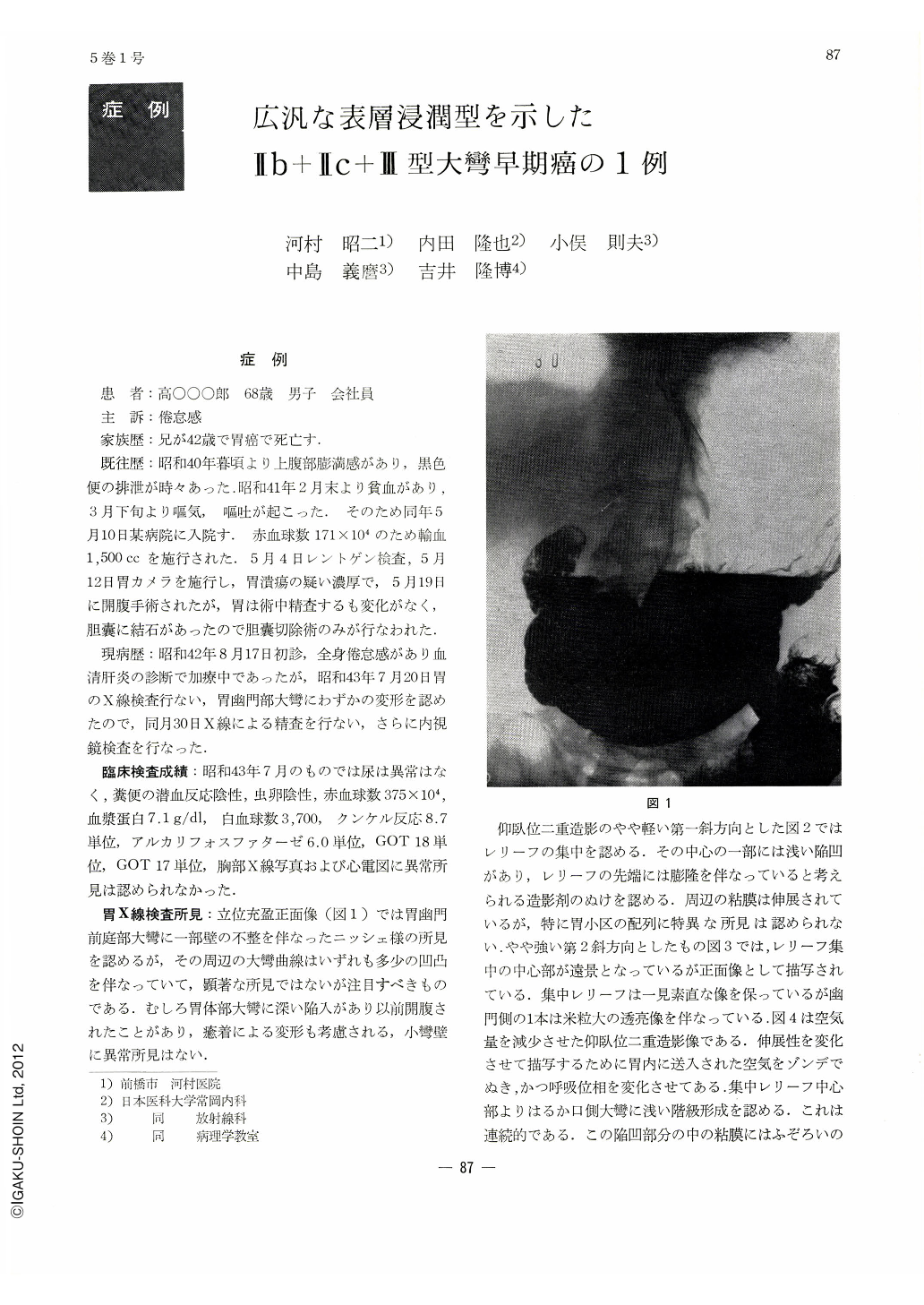
X-ray examination: Slight rigidity was noticed in upright barium filled pictures on the greater curvaure of the gastric antrum, where mucosal convergence was visualized both in double contrast and compression studies. Continuous grade-formation such as swelling and narrowing of the central portion clearly indicated the presence of a small Ⅱc+Ⅲ. However, in pictures in which the pliability of the gastric wall was retained, there was noticed in an oral area away from the center a step-formation as of a distinct hyperbolic curve, and granular formation was recognized in the mucosa in this shallow and wide depression. Irregular, abnormal areas were also noticed in regions outside the grade-formation. It was considered then that cancer was not restricted within the central part of the mucosal convergence but was more extensive; its sphere remained uncertain, however. Endoscopically biopsy was positive for cancer in all specimens taken from areas of engorgement, swelling and white exudate in the center of mucosal convergence.
Gross and histological findings: A depression with Ul-Ⅱ mucosal convergence was seen on the greater curvature of the antrum. Its niveau was clearly on a different level from the surrounding areas associated with granular formation. In an area near the lesser curvature was recognized partly a stair-like like formation as of Ⅱc. Histologically the central part was Ⅱc accompanied with ulcer scar, and the surrounding parts were mostly Ⅱb with cancer invasion in the upper half of the mucosal layer, with only a slight part of it being Ⅱc. As a whole it was adenocarcinoma tubulare, measuring 9 by 6cm.
In x-ray and endoscopical examinations of early gastric cancer, especially in a case like this, extensive Ⅱb with Ⅱc+Ⅲ, it is not only necessary to notice the different levels of mucosal niveau and changes of mucosal hue but also it is essential, for confirmation of the extent of cancer, to discriminate the normal mucosa from the malignant one by means of biopsy.

Copyright © 1970, Igaku-Shoin Ltd. All rights reserved.


