Japanese
English
- 有料閲覧
- Abstract 文献概要
- 1ページ目 Look Inside
症例
患者:45歳 男
既往歴:家族歴に特記すべき所見なし,
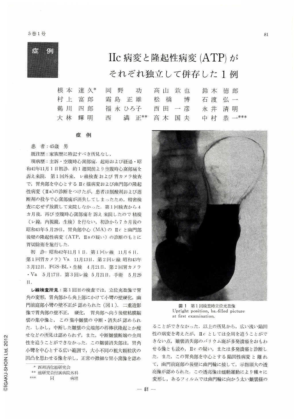
現病歴:主訴・空腹時心窩部痛.起始および経過・昭和42年11月1日初診.約1週間前より空腹時心窩部痛を訴え来院.第1回外来,レ線検査および胃カメラ検査で,胃角部を中心とするⅡc様病変および幽門部の隆起性病変(Ⅱa)の診断をつけたが,患者は制酸剤および遮断剤の投与で心窩部痛が消失してしまったため,精密検査に応ぜず放置して来院しなかった.第1回検査から4カ月後,再び空腹時心窩部痛を訴え来院したので精検(レ線,内視鏡,生検)を行ない,初診から7カ月後の昭和43年5月29日,胃角部中心(MA)のⅡcと幽門部後壁の隆起性病変(ATP,Ⅱaの疑い)の診断のもとに胃切除術を施行した.
This is a case report of a 45-year-old man visiting the authors' hospital with hunger pain as his chief complaint. In his stomach was found a wide Ⅱc with the gastric angle as its center, coexistent with another polypoid lesion (Ⅱa-like aggregate of atypical epithelium) on the posterior wall of the antrum adjacent to the pyloric ring. They were located at some distance from, and independent of, each other
At the initial examinations of x-ray and endoscopy, he was suspected as having Ⅱc or multiple ulcers coexistent with Ⅱa lesion. However, as his chief complaint disappeared by the administration of antiacid agents, further examination was neglected for 3 or 4 months. Surgical intervention was eventually performed 7 months later from the intial examination.
It was early gastric cancer with the degree of depth invasion luckily localized mostly within the “pm”, with only a small part of it reaching the “sm”. No metastasis was found as far as the secondary lymph nodes (no, 0/33).
This case is of interest from the following points: ――
(1) Both roentgenologically and endoscopically, preoperative diagnosis was diflicult to make as to whether Ⅱc and polypoid lesion were independent of each other; conversely, it was hard to determine the extent of Ⅱc lesion, especially its pyloric border.
(2) In x-ray pictures, barium fleck over the area, where the gastric rugae were about to disappear, was of variegated shape so that multiple ulcers were suspected as well. Until the result of fibergastroscopic biopsy was made known, reactive lymporeticular hyperplasia had also been suspected as the extent of Ⅱc was impossible to determine.
(3) The protrusion in the pyloric part was diagnosed as ATP by biopsy, but by both x-ray and endoscopy it was definitely and indubitably regarded as Ⅱa. Such a lesion as exists on the border area between benign and malignant lesions should be clinically dealt with at present as of malignant nature.
Copyright © 1970, Igaku-Shoin Ltd. All rights reserved.