Japanese
English
- 有料閲覧
- Abstract 文献概要
- 1ページ目 Look Inside
- サイト内被引用 Cited by
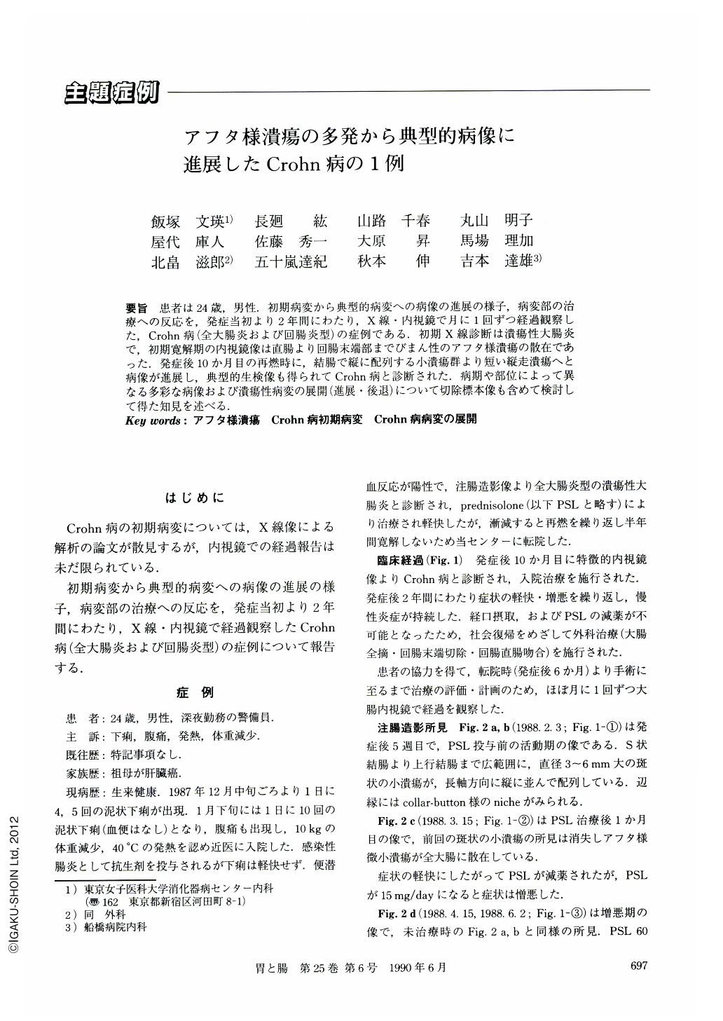
要旨 患者は24歳,男性.初期病変から典型的病変への病像の進展の様子,病変部の治療への反応を,発症当初より2年間にわたり,X線・内視鏡で月に1回ずつ経過観察した,Crohn病(全大腸炎および回腸炎型)の症例である.初期X線診断は潰瘍性大腸炎で,初期寛解期の内視鏡像は直腸より回腸末端部までびまん性のアフタ様潰瘍の散在であった.発症後10か月目の再燃時に,結腸で縦に配列する小潰瘍群より短い縦走潰瘍へと病像が進展し,典型的生検像も得られてCrohn病と診断された.病期や部位によって異なる多彩な病像および潰瘍性病変の展開(進展・後退)について切除標本像も含めて検討して得た知見を述べる.
A 24-year-old male with Crohn's colitis, diffuse type, was followed up by double contrast barium enema and colonoscopy, about once a month for two years. The course of evolution and retrogression of the ulceration, and polymorphous features according to the stage and distribution of the lesions are described in this report (Figs. 12 and 13). Involved areas were the total colon, rectum, and the terminal ileum. Anal lesions and other complications were not found. After medication with prednisolone and nutritional treatment in hospital for 2 years, resection of the total colon and the terminal ileum, and recto-ileostomy were done because of chronic inflammation.
Numerous aphthoid ulcers were scattered on the normal mucosa throughout the entire colon and the terminal ileum. These were seen in the clinically remitting period at the early stage (Figs. 2 b, 3 a, b) and progressed to the typical lesions of Crohn's disease at ten months after the onset.
Evolution of the ulceration occurred as follows; discrete ulcers were first arranged on a few longitudinal lines (Fig. 4), fusion of these ulcers then occurred resulting in the typical longitudinal ulcers in the same sites (Fig. 5).
On the other hand, the lesion of these discrete ulcers retrogressed to the lesion of the scattered aphthoid ulcers twice in the clinically remitting period in six months after the onset.
New aphthoid ulcers were found on the marginal mucosa of the longitudinal ulcers or ulcer scars (Fig. 10 c), which were elevated by underlying inflammation and edema in the submucosa, in the resected specimen (Fig. 11). They were arranged in a longitudinal direction, too.

Copyright © 1990, Igaku-Shoin Ltd. All rights reserved.


