Japanese
English
- 有料閲覧
- Abstract 文献概要
- 1ページ目 Look Inside
Crohn病は慢性に進行する消化管の炎症と言われ,腸管壁の肥厚,線維化などを伴いやすいため,その合併症としては瘻孔形成や狭窄の頻度が高く,腹腔内への穿孔はまれと考えられてきた.しかし近年内外で穿孔例の報告が増え,Crohn病の急性増悪に伴う合併症とする考え方もみられる.われわれも回腸にfree perforationを起こしたCrohn病の1例を経験したので報告し,穿孔の原因などについて若干の考察を試みる.
症 例
患 者:23歳,男性.
主 訴:腹痛,腹部膨満感.
家族歴:父,急性肝炎.
既往歴:20歳時,痔瘻の手術.
現病歴:1年前より上腹部痛があり,近医で胃内視鏡検査を受け胃多発ポリープの診断および3カ月後の再検査で縮小していると言われた.1978年10月ごろより臍周囲から右下腹部を中心とする鋭い痂痛と腹鳴を時々認めるようになったが,悪心,嘔吐,発熱はなかった.1979年になり腹部膨満感,食欲不振,体重減少も加わり当院受診し,同年5月7日精査のため入院となった.
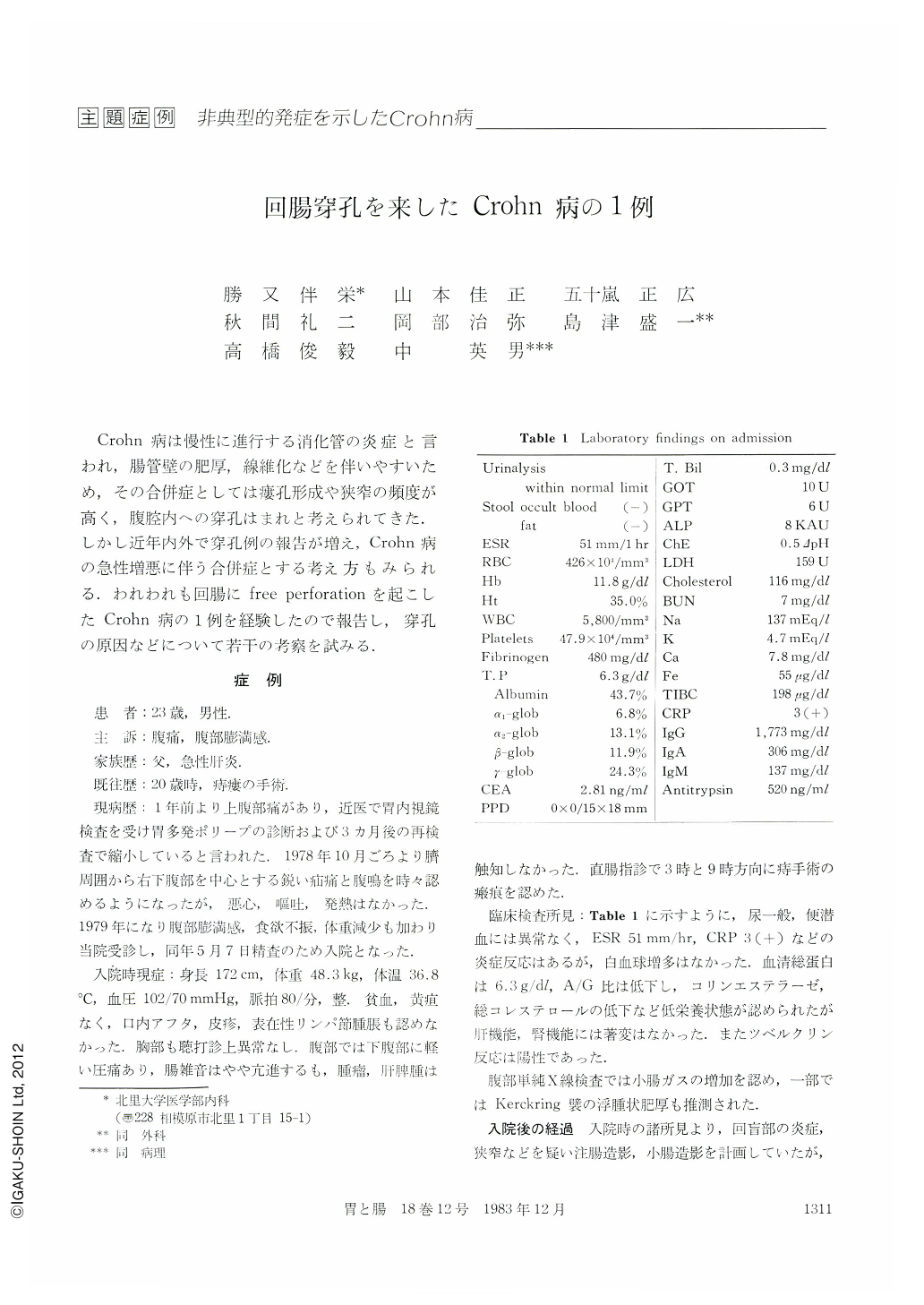
A 23-year-old man was admitted to the Kitasato University Hospital with a 6-month history of right lower quadrant pain, weight loss and abdominal fullness on May 7, 1979. On admission, he was afebrile with slight abdominal tenderness but no abdominal mass was palpable. The hemoglobin was 11.8 g per 100 ml, white blood cell count was 5,800, serum albumin was 2.7 g per 100 ml, CRP was 3 positive. Abdominal x-ray pictures showed increased gas shadow of the small bowel.
On the 4th day after admission, he developed severe abdominal pain and marked distension with high fever (38.8℃). Physical examination revealed abdominal guarding and rebound tenderness. Emergency exploratory abdominal laparotomy was performed with a diagnosis of acute peritonitis. At laparotomy, large amount of purulent fluid was found in the peritoneal cavity. The distal ileum and cecum were indurated and congested markedly. A small perforation was seen on the antimesenteric border of the ileum about 30 cm proximal to the ileocecal valve. After resection of approximately 100 cm of the ileum, the cecum and 12 cm of ascending colon, a jejuno-ascending colostomy was constructed.
Macroscopic findings of the resected specimen showed typical regional enteritis. There were three narrow segments associated with longitudinal ulcers and cobblestone appearances. Microscopically, the area of the cobblestones showed transmural inflammation accompanied by multiple fissuring ulcers and abscess formations.
After operation, the patient was treated with salazosulfapyridine and remained asymptomatic for about four years. A small bowel series revealed relapsing of regional enteritis on June, 1983. Multiple lineal ulcers and pseudodiverticular formations are seen from the anastomotic area to the jejunum.

Copyright © 1983, Igaku-Shoin Ltd. All rights reserved.


