Japanese
English
- 有料閲覧
- Abstract 文献概要
- 1ページ目 Look Inside
- サイト内被引用 Cited by
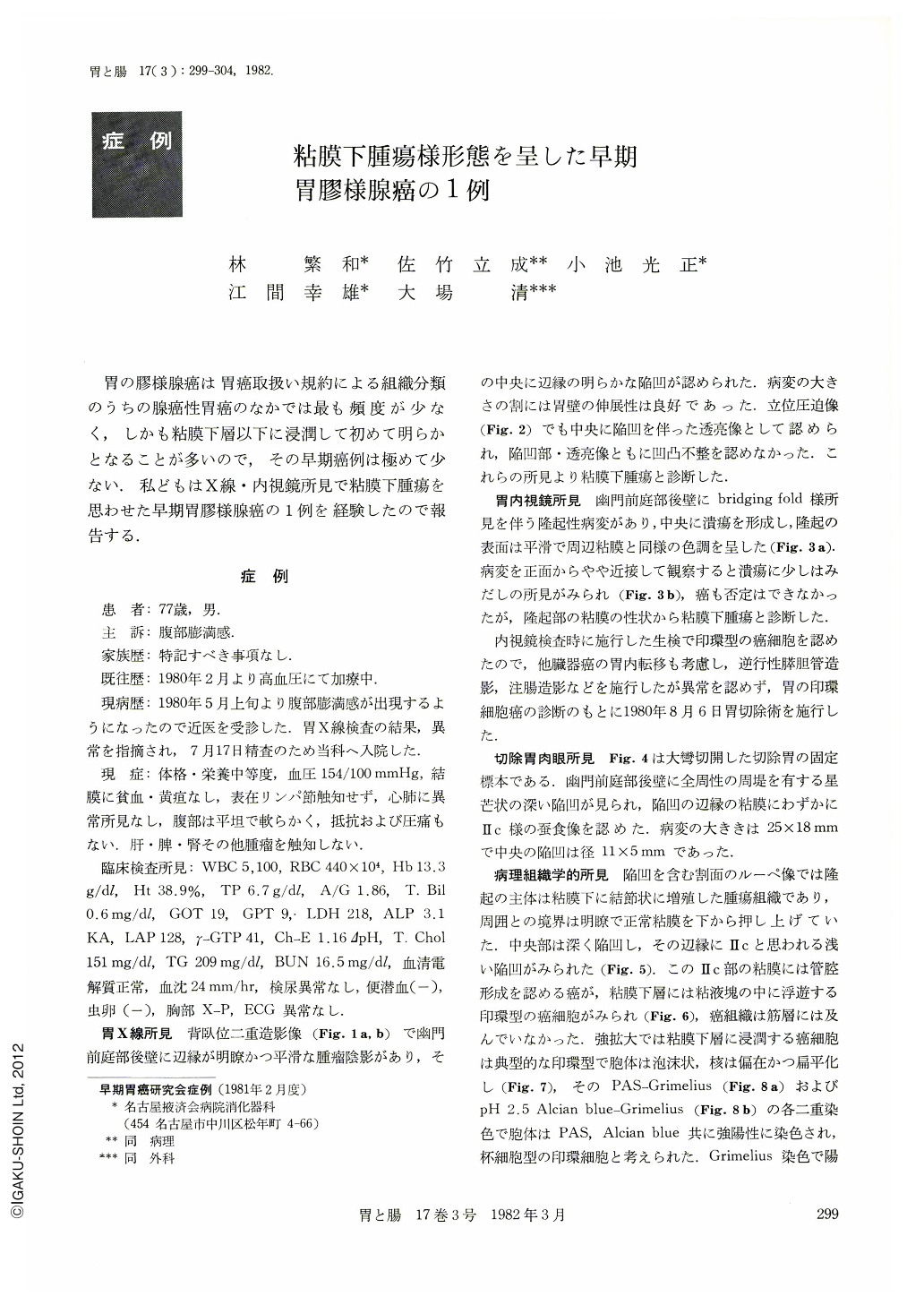
胃の膠様腺癌は胃癌取扱い規約による組織分類のうちの腺癌性胃癌のなかでは最も頻度が少なく,しかも粘膜下層以下に浸潤して初めて明らかとなることが多いので,その早期癌例は極めて少ない.私どもはX線・内視鏡所見で粘膜下腫瘍を思わせた早期胃膠様腺癌の1例を経験したので報告する.
The patient: a man aged 77. X-ray and endoscopic examination of the stomach showed on the posterior wall of the antrum a smooth, sharply circumscribed elevated lesion accompanied with depression. It was suggestive of a submucosal tumor, but as biopsy revealed malignant cells of signet-ring type, we took also into account some metastatic carcinoma. However, as examinations of the other organs showed no malignant findings, the patient was operated on under a diagnosis of gastric carcinoma. The resected specimen showed an elevated lesion, measuring 25×18 mm, with a deep depression in the center, measuring 11×5 mm. Slight Ⅱc-like encroachment was seen in the margins of the depression. Histologically, the chief part of the elevation was tumor tissue in the submucosa showing nodular proliferation, forcing up the normal mucosa from underneath. Around the borders of the deep excavation were seen Ⅱc-like changes, showing tubular adenocarcinoma. Cancer cells of signet-ring type were seen floating in the mucous lake in the submucosa. As the cell body showed marked positive reaction to PAS and Alcian blue, the cells were considered to belong to signet ring type cancer cells of goblet cell type. The diagnosis was poorly differentiated mucinous adenocarcinoma with depth of invasion reaching the submucosa.
Mucinous adenocarcinoma is least frequent among adenocarcinomas of the stomach, and its histological type is confirmed only when it proliferates beneath the submucosa. Accordingly, early gastric mucinous adenocarcinoma as in the present case localized within the submucosa is of rare occurrence.
Diagnosis was difficult because it had ample features of radiographical and endoscopic findings to warrant a submucosal tumor. One should take into account the possibility of mucinous adenocarcinoma when such a shape of cancer be encountered and signet-ring type cells be demonstrated by biopsy.

Copyright © 1982, Igaku-Shoin Ltd. All rights reserved.


