Japanese
English
- 有料閲覧
- Abstract 文献概要
- 1ページ目 Look Inside
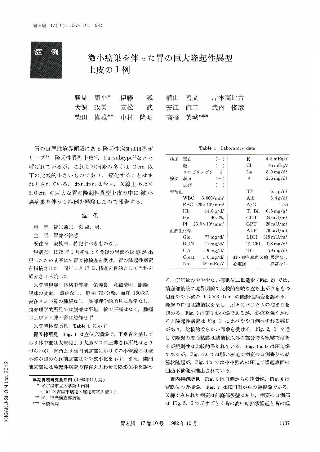
胃の良悪性境界領域にある隆起性病変はIII型ポリープ2),隆起性異型上皮4),IIa-subtype5)などと呼ばれているが,これらの病変の多くは2cm以下の比較的小さいものであり,癌化することはまれとされている.われわれは今回,X線上6.5×3.0cmの巨大な胃の隆起性異型上皮の中に微小癌病巣を伴う1症例を経験したので報告する.
A 61-year-old man visited a neighboring doctor with a complaint of epigastric discomfort and gastric polypoid lesion was found by x-ray examination. On January 17, 1978, he was admitted to our hospital for detail examination.
X-ray examination of the stomach revealed a large polypoid lesion which measured 6.5×3.0 cm in the posterior wall of the antrum. The surface of the lesion was uneven and mucosal pattern was rough, which means the lesion was epithelial origin. Although the mucosal pattern of the lesion was rough, areae gastricae was rather kept in regular manner. And no ulceration was seen. In both double contrast and compression study, the lesion changed its shape a little by changing photographing position, which means the lesion was rather soft.
Endoscopic examination revealed a large protruded lesion which was slightly white in the posterior wall of the antrum. There was neither redness nor ulceration in the surface of the lesion. Although x-ray and endoscopic findings had the features of atypical epithelium, the lesion was diagnosed as early type I carcinoma because of inordinately large lesion. All six biopsy specimens were diagnosed as Group III. And we finally diagnosed most of this lesion to atypical epithelium.
Considering the possibility of focal carcinoma, operation was done on Junuary 31, 1978. A flower-bed like flat protruded lesion was recognized in the posterior wall of the antrum and there was neither redness nor ulceration in the surface of the lesion.
Histological study revealed atypical glands in the surface of the mucosa and cystic dilatation of glands in the deeper layer. Although most of the lesion was diagnosed as atypical epithelium, and minute cancerous lesions were observed partly.
It seemed to be important to make it appear by x-ray and endoscopic examination for making the diagnosis of atypical epithelium that the lesion has a rather regular mucosal pattern in the surface, a rather soft nature and has a somewhat white surface, which are distinguishable points from protruded type carcinoma. Although atypical epithelium is the lesion which permits follow-up study, large lesions more than 4 cm in diameter should be operated on because of the highly possible focal carcinoma.

Copyright © 1982, Igaku-Shoin Ltd. All rights reserved.


