Japanese
English
- 有料閲覧
- Abstract 文献概要
- 1ページ目 Look Inside
最近,われわれは数個の小さな粘膜隆起が2年5カ月間に著明な変化を示し,巨大皺襞所見を呈した進行胃悪性リンパ腫を経験したので報告する.
症 例
患 者:51歳,男,会社員.
主 訴:食思不振,心窩部痛.
家族歴:特記することなし.
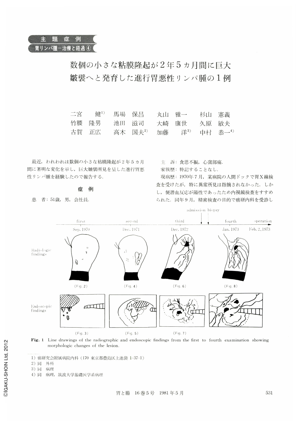
現病歴:1970年7月,某病院の人間ドックで胃X線検査を受けたが,特に異常所見は指摘されなかった.しかし,便潜血反応が陽性であったため内視鏡検査をすすめられた.同年9月,精密検査の目的で癌研内科を受診した.胃X線(9月10日)および内視鏡検査(9月12日)では,異常なしと診断された.1971年12月4日(1年3ヵ月後)のX線検査では,胃体上部の粘膜下腫瘍と診断され,内視鏡検査(12月5日)では,同部位の限局性肥厚性胃炎と診断された.患者は,1972年8月ごろより食思不振と心窩部膨満感を覚えるようになり,同年9月20日,某病院で胃X線検査を受けたが異常所見は指摘されなかった.しかし上記症状は改善せず,同年12月中句ごろよりとう痛を伴うようになったため,同年12月25日(2回目の検査の1年後)再び癌研内科を受診.胃X線検査(12月25日)の結果,胃体上部の進行癌と診断され,1973年1月19日手術の目的で入院した.
A 51-year-old male patient was admitted to the Dept. of Internal Medicine, Cancer Institute Hospital, Tokyo, on January 19, 1973, under the diagnosis of malignant lymphoma of the stomach. His chief complaint was anorexia and epigastric pain.
In June 1970 he received health check, including upper G-I series. The stomach was reported to be normal. But endoscopy was recommended because of positive occult blood test. In September 1970 he had the first visit to the Dept. of Internal Medicine, C.I.H. for the detailed examination. The first x-ray (Sept. 10, 1970) and endoscopy (Sept. 12, 1970) were reported to be normal. The second x-ray done on December 4, 1971, revealed the localized thickening of the mucosa in the upper gastric body, and the diagnosis of submucosal tumor was made. But in the second endoscopy the abnormality detected by the x-ray was merely diagnosed as localized chronic hypertrophic gastritis. No further study had been done until August 1972 when he noticed anorexia and epigastric fullness. In September 1972 x-ray of the stomach was done in an other hospital. But no abnormality was detected.
He had the third visit to the C.I.H. on December 25, 1972, because of epigastric pain which had appeared in the middle of December. The third x-ray of the stomach done on December 25, 1972, revealed markedly enlarged folds of the fornix and middle body which were strongly suggestive of malignancy.
Physical examination on admission was normal except for slight tenderness in the upper epigastrium. Blood count, blood chemistry and urinalysis were normal. The fourth x-ray and endoscopy suggested malignant lymphoma and it was confirmed by endoscopic biopsy. Operation (total gastrectomy with combined resection of the distal esophagus and the tail of the pancreas, curative) was done on February 2,1973. Histologic examination of the resected material revealed malignant lymphoma, diffuse histiocytic type with extensive involvement of the mucosa and submucosa. and slight involvement of the propria muscle and subserosa. No metastasis was found in the dissected lymph nodes (0/23).

Copyright © 1981, Igaku-Shoin Ltd. All rights reserved.


