Japanese
English
- 有料閲覧
- Abstract 文献概要
- 1ページ目 Look Inside
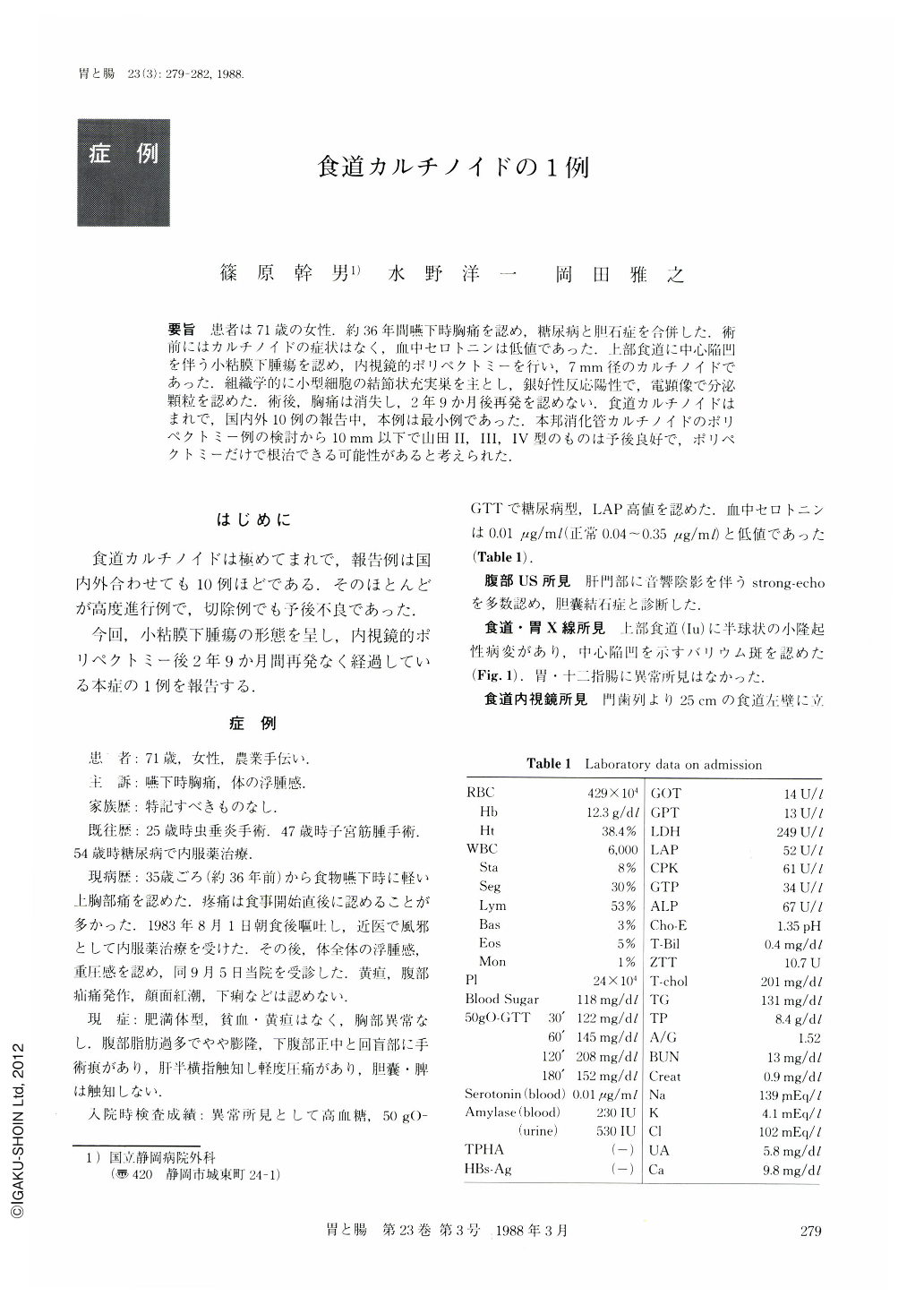
要旨 患者は71歳の女性.約36年間嚥下時胸痛を認め,糖尿病と胆石症を合併した.術前にはカルチノイドの症状はなく,血中セロトニンは低値であった.上部食道に中心陥凹を伴う小粘膜下腫瘍を認め,内視鏡的ポリペクトミーを行い,7mm径のカルチノイドであった.組織学的に小型細胞の結節状充実巣を主とし,銀好性反応陽性で,電顕像で分泌顆粒を認めた.術後,胸痛は消失し,2年9か月後再発を認めない.食道カルチノイドはまれで,国内外10例の報告中,本例は最小例であった.本邦消化管カルチノイドのポリペクトミー例の検討から10mm以下で山田Ⅱ,Ⅲ,Ⅳ型のものは予後良好で,ポリペクトミーだけで根治できる可能性があると考えられた.
A 71 year-old woman, whose health was complicated by diabetes mellitus and cholelithiasis, had had a long history of chest pain on swallowing. The patient had no carcinoid symptoms and laboratory data showed a low level of serotonin in the blood. X-ray and endoscopic examination of the esophagus disclosed a small submucosal tumor with a shallow depression on its top in the upper esophagus (Fig. 1, 2), which was resected by endoscopic polypectomy. The tumor was 7 mm in diameter and diagnosed histologically as carcinoid, showing positive argyrophil reaction (Fig. 3, 4) . Postoperatively, the patient has been well 2 years and 9 months, with neither chest pain nor any finding of recurrence of the tumor.
Ten reported cases of carcinoid of the esophagus were reviewed (Table 2), and discussed regarding gross pictures, histological findings and the prognosis.
An analysis of polypectomy cases in Japan revealed the possibility of radical treatment by endoscopic polypectomy in cases where the lesion is less than 10 mm in diameter and Yamada's type Ⅱ, Ⅲ, Ⅳ in shape.

Copyright © 1988, Igaku-Shoin Ltd. All rights reserved.


