Japanese
English
- 有料閲覧
- Abstract 文献概要
- 1ページ目 Look Inside
- サイト内被引用 Cited by
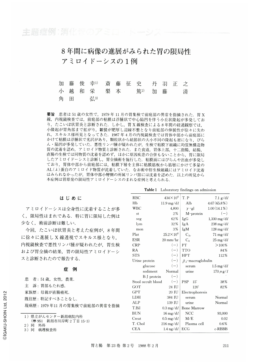
要旨 患者は51歳の女性で,1979年11月の胃集検で前庭部の異常を指摘された.胃X線,内視鏡検査では,前庭部の粘膜は浮腫状で中心陥凹を伴う小丘状隆起が多発しており,たこいぼ状胃炎と診断された.しかし,胃X線検査による8年間の経過観察では,小隆起が胃角部まで拡がり,皺襞が肥厚し辺縁不整となり前庭部の伸展性が徐々に失われ,スキルス様所見となってきた.1987年4月の内視鏡検査では胃体中部から前庭部にかけて粘膜は浮腫状で光沢があり,顆粒状から結節状の大小不同の隆起も密になり,びらん・陥凹が多発していた.悪性リンパ腫が疑われたが,生検で粘膜下組織に均質無構造物質の沈着を認め,アミロイド物質と診断された.また食道,胃体上部,十二指腸,結腸,直腸の生検では同物質の沈着を認めず,ほかに原因疾患の合併もないことから,胃に限局したアミロイドーシスと診断し,胃全摘術を施行した.粘膜面にはびらんや出血が多発しており,胃体中部から前庭部には,粘膜下層を主体に粘膜筋板から筋層にかけて多量のAL(λ)蛋白のアミロイド物質が沈着していた.なお術中肝生検組織にはアミロイド沈着はみられなかったが,胃体中部小彎側の所属リンパ節には沈着を認めた.以上の所見から本症例は胃原発の限局性アミロイドーシスのまれな症例と考えられる.
A 51-year-old woman was found to have abnormality in the gastric antrum by mass screening and diagnosed as having erosive gastritis endoscopically in Dec. 1979. Follow-up study of upper GI series performed in April 1987 showed polypoid lesions and thickened mucosa suggesting scirrhous type carcinoma. Subsequently performed endoscopic examination revealed thickened elastic mucosa with slight discoloration, irregularly elevated mocosa and multiple erosion. Biopsy specimens showed amyloid in the gastric submucosa. Biopsies of other places of the GI tract, however, were all negative for amyloid deposition.
Total gastrectomy was performed in July 1987 because of mucosal friability and progressive lesions. In the resected stomach, amyloid (light chain protein λ) was observed in the antrum and the lower portion of the body. Amyloid was also found in the regional lymph nodes along the lesser curvature of the stomach. Liver biopsy performed during operation was negative for amyloid.
Based on these findings, this case was diagnosed as localized primary amyloidosis of the stomach.

Copyright © 1988, Igaku-Shoin Ltd. All rights reserved.


