Japanese
English
- 有料閲覧
- Abstract 文献概要
- 1ページ目 Look Inside
ヒト胃癌モデルとしてイヌ実験胃癌を研究するに当っては,いかにしてヒト胃癌に似たイヌ進行胃癌を作るかに精力が注がれてきた1)~5).そして今やイヌ胃癌は,肉眼型も組織型もヒト胃癌に似て多彩で,リンパ節転移,肝,肺,骨など臓器転移,皮膚転移,癌性腹膜炎の併発まで作られている4)5).MNNGやENNG単独投与では極めて稀なイヌスキルスの発生も,ガストリンを併用すると意図的に作れる段階にきている6).さらに実験癌が悪性病変であるとの証明に転移形成と共に移植継代が必要とされるが,ヌードマウスを用いて成功している7)8).
このような背景の下にイヌ実験胃癌を用いる微小癌研究の意義は,①意図的に微小癌を作ってその切除胃の詳細な検索で組織発生を追求する,②内視鏡下の生検で確認された微小癌が増殖,発育して進行癌へと進展して行く過程をX線,内視鏡,生検で経時的に詳細な追求をすることにあろう.こうした微小癌に的をしぼった研究の他に,ヒト胃癌で切除胃の全割標本に微小癌がみつけられた全く同じ方法をイヌ実験胃癌にも当てはめてみるのもヒト胃癌の対比に役立つと考えられる.
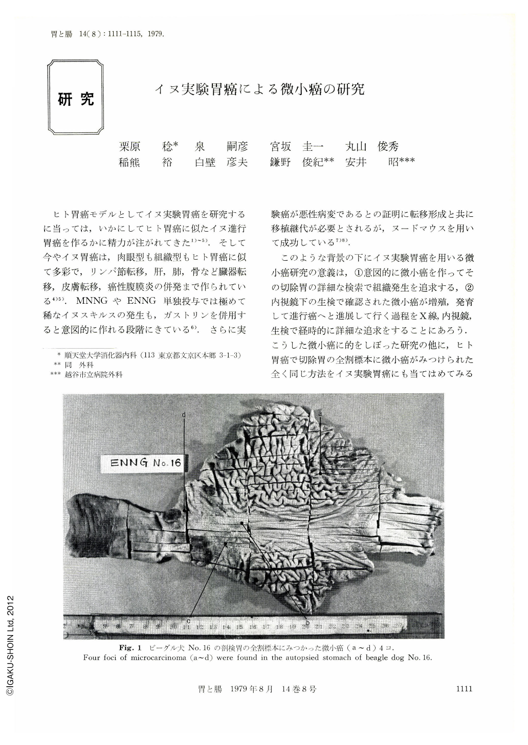
By making 5mm-width section of the canine stomach, 33 cases of ENNG or MNNG induced canine gastric cancer were investigated at four “institutions (Juntendo Univ., Tokyo Medical and Dental Univ. (Murakami), Kyushu Univ. (Kodama), Osaka Univ. (Fujita)). Then we found 96 minute gastric cancers whose diameter were less than 5mm among them, 56 lesions were located at the upper portion of the stomach (C), 16 lesions were at the middle portion of the stomach (M) and 24 lesions were at the lower portion of the stomach (A).
Similar to the advanced gastric cancer, their distribution was particularly predominant at anterior wall and greater curvature of the body of the stomach as well as antrum. Macroscopically, 4 lesions were type I, 16 lesions were type Ⅱa, 35 lesions were type Ⅲb, 39 lesions type Ⅱc and 2 lesions were type Ⅲ.
Their depth of invasion showed that 83 lesions were limited in mucosa (M) and 13 lesions were down to the submucosa (sm). Histologic findings were as follows;
Among the 45 minute gastric cancers which were induced by Tween 60 plus ENNG, well-differentiated type was more dominant than undifferentiated type in the upper portion of the stomach (C), and undifferentiated type was more prominent in the lower portion of the stomach (A). On the other hand, in 51 lesions of MNNG induced minute gastric cancer, differentiated cancer was 86.5% in the upper portion of the stomach (C) and 91.7% in the middle portion of the stomach.
In experimental canine gastric cancer, it is possible to follow the development and progression of the cancer by doing X-ray study, endoscopy and biopsy. In months after starting administration of ENNG, discoloration of gastric mucosa, loss of mucosal luster and granular change of mucosa developed. In 6 months later, multiple tiny errosive mucosal lesions occurred and approximately 1 year after the administration of ENNG, minute gastric cancer developed and it could be diagnosed by biopsies.
Therefore, as Murakami tried, we can study the development of the minute gastric cancer by resecting this stage of the experimental canine gastric cancer. As mentioned previously, we can closely persue the developing process of the minute gastric cancer and it's progression into the advanced cancer in the canine stomach by using X-ray and endoscopy.
Minute gastric cancer gradualy develops, then forms multiple small nodular lesions and finally they fuse into a prominent protruded cancer. On the other hand, they may become Borr. type 3 cancer through the malignant cycle which is commonly seen in human being.
Copyright © 1979, Igaku-Shoin Ltd. All rights reserved.