Japanese
English
- 有料閲覧
- Abstract 文献概要
- 1ページ目 Look Inside
陥凹型微小胃癌のうち術前に確診しえた典型的症例を示し,併せて若干X線診断の問題点について述べたい.
症例
患者:N. C. 40歳 男性
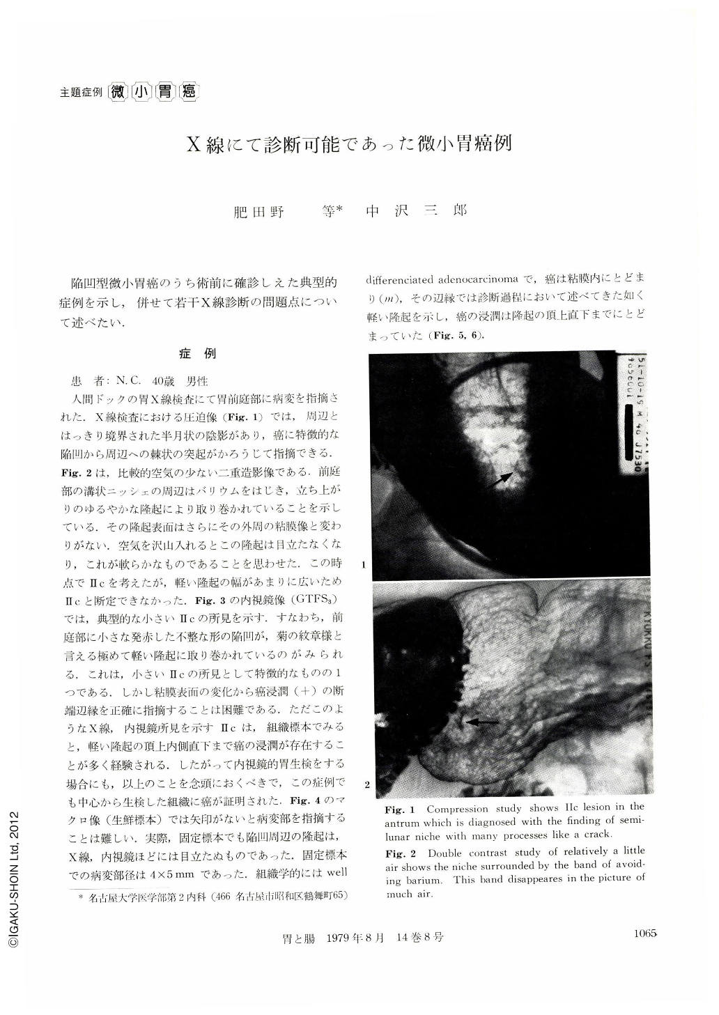
入間ドックの胃X線検査にて胃前庭部に病変を指摘された.X線検査における圧迫像(Fig. 1)では,周辺とはっきり境界された半月状の陰影があり,癌に特徴的な陥凹から周辺への棘状の突起がかろうじて指摘できる.Fig. 2は,比較的空気の少ない二重造影像である.前庭部の溝状ニッシェの周辺はバリウムをはじき,立ち上がりのゆるやかな隆起により取り巻かれていることを示している.その隆起表面はさらにその外周の粘膜像と変わりがない.空気を沢山入れるとこの隆起は目立たなくなり,これが軟らかなものであることを思わせた.この時点でⅡcを考えたが,軽い隆起の幅があまりに広いためⅡcと断定できなかった.Fig. 3の内視鏡像(GTFS3)では,典型的な小さいⅡcの所見を示す.すなわち,前庭部に小さな発赤した不整な形の陥凹が,菊の紋章様と言える極めて軽い隆起に取り巻かれているのがみられる.これは,小さいⅡcの所見として特徴的なものの1つである.しかし粘膜表面の変化から癌浸潤(+)の断端辺縁を正確に指摘することは困難である.ただこのようなX線,内視鏡所見を示すⅡcは,組織標本でみると,軽い隆起の頂上内側直下まで癌の浸潤が存在することが多く経験される.したがって内視鏡的胃生検をする場合にも,以上のことを念頭におくべきで,この症例でも中心から生検した組織に癌が証明された.Fig. 4のマクロ像(生鮮標本)では矢印がないと病変部を指摘することは難しい.実際,固定標本でも陥凹周辺の隆起は,X線,内視鏡ほどには目立たぬものであった.固定標本での病変部径は4×5mmであった.組織学的にはwell diiferenciated adenocarcinomaで,癌は粘膜内にとどまり(m),その辺縁では診断過程において述べてきた如く軽い隆起を示し,癌の浸潤は隆起の頂上直下までにとどまっていた(Fig. 5, 6).
In the diagnosis of gastric cancer one of the most important factors is whether or not we are able to determine the vertical changes with the mucosal surface as their border, or the degree of unevenness of the mucosal surface, and horizontal changes on the mucosal surface, or the spread of cancer. Early cancer with no changes of the former is Ⅱb, and slight changes in the latter is microcarcinoma. Both are hard to diagnose by X-ray. Diagnosis of microcarcinoma of Ⅱb type would remain most difficult. Most of relatively extended Ⅱb hitherto reported show some, even if slight, unevenness of the mucosal surface. Therefore, diagnosis of minute Ⅱc or Ⅱa type is a good match for that of extended Ⅱb type in its difiiculty. Even in microcarcinoma of Ⅱe type less than 5mm indiameter some have changes in the surrounding area and some have none at all. Ⅱc type microcarcinomas being detected or correctly diagnosed by us are only those that belong to the former. The case here presented is their typical one. A small lesion, measuring 0.4×0.5cm, located on the posterior wall of the gastric antrum, was found in a man aged 40. Macroscopically it was not distinct in the excised speciman, but in and biopsy examinations slight, non-cancerous elevation was recognized around the minute cancer. Such a finding is typical for microcarcinoma of this size. Correct diagnosis was obtained by moderate compression in X-ray study. Taking advantage of such non-cancerous changes can facilitate the detection on diagnosis of minute carcinoma. Obstacles in diagnosis sometimes depend on the location of cancer.
At all events, clues to the diagnosis of incipient carcinoma should cover not only horizontal diameters of cancer but also three dimentional consideration in diagnosis.

Copyright © 1979, Igaku-Shoin Ltd. All rights reserved.


