Japanese
English
- 有料閲覧
- Abstract 文献概要
- 1ページ目 Look Inside
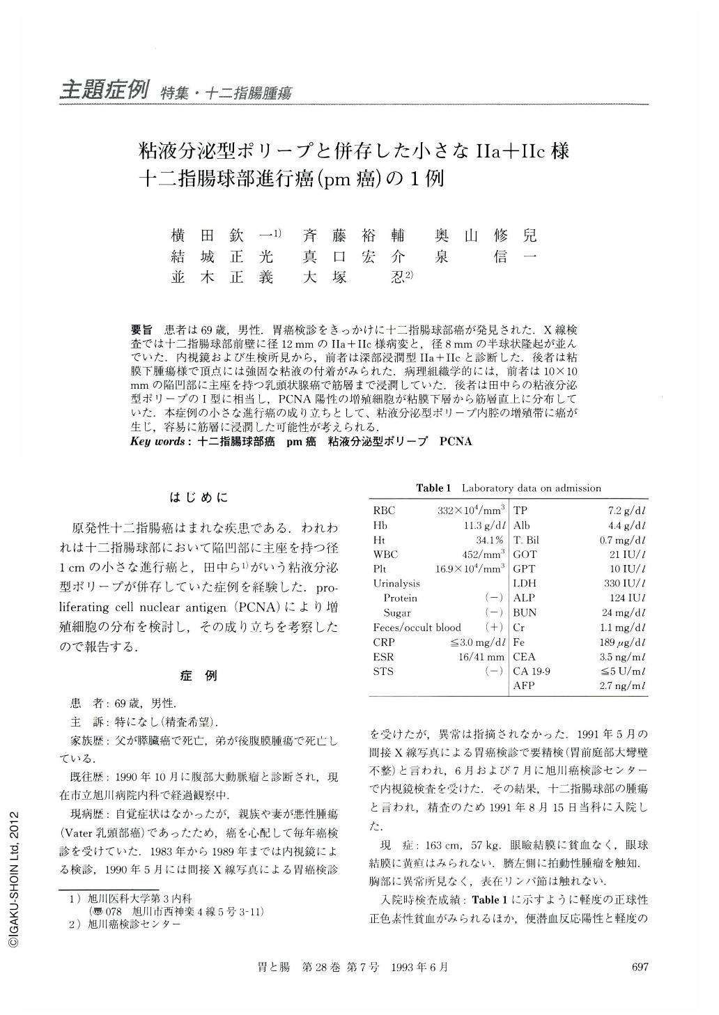
要旨 患者は69歳,男性.胃癌検診をきっかけに十二指腸球部癌が発見された.X線検査では十二指腸球部前壁に径12mmのⅡa+Ⅱc様病変と,径8mmの半球状隆起が並んでいた.内視鏡および生検所見から,前者は深部浸潤型Ⅱa+Ⅱcと診断した.後者は粘膜下腫瘍様で頂点には強固な粘液の付着がみられた.病理組織学的には,前者は10×10mmの陥凹部に主座を持つ乳頭状腺癌で筋層まで浸潤していた.後者は田中らの粘液分泌型ポリープの1型に相当し,PCNA陽性の増殖細胞が粘膜下層から筋層直上に分布していた.本症例の小さな進行癌の成り立ちとして,粘液分泌型ポリープ内腔の増殖帯に癌が生じ,容易に筋層に浸潤した可能性が考えられる.
A 69-year-old man was admitted to our hospital for a further evaluation of a duodenal cancer. Screening x-ray examination for stomach cancer revealed antral deformity. More detailed upper gastrointestinal x-ray and endoscopic examination showed a small type Ⅱa+Ⅱc like lesion and a hemispherical polyp on the anterior wall of the duodenal bulb. The latter polyp secreted mucus from an orifice. The biopsy specimen from the former lesion disclosed a well-differentiated adenocarcinoma. Resected specimen showed an irregularly depressed lesion with marginal elevation, measuring 10×10 mm in size, and a hemispherical polyp, measuring 6×6 mm in size, in the duodenal bulb. Histologically the former was a papillary adenocarcinoma invading the muscular layer, and the latter was consistent with a mucus-secretingpolyp, type Ⅰ being reported by Tanaka et al. Epithelium of this polyp was invaginated into the submucosal layer from an orifice, resulting in the formation of a lumen, whose wall was composed of gastric type foveolar epithelium and Brunner's glands. There was proliferating cell nuclear antigen along the boundary between gastric type villi and Brunner's glands. In the bottom of the polyp proliferating cells formed a line just above the muscular layer. Papillary adenocarcinoma of our case did not have brush border and goblet formation, which suggested gastric differentiation. Malignant transformation seemed to arise from the proliferating zone of mucus-secreting polyps and invade the muscular layer easily. Small advanced cancer, less than 10 mm in size, of the duodenum is rare in the literature.

Copyright © 1993, Igaku-Shoin Ltd. All rights reserved.


