Japanese
English
- 有料閲覧
- Abstract 文献概要
- 1ページ目 Look Inside
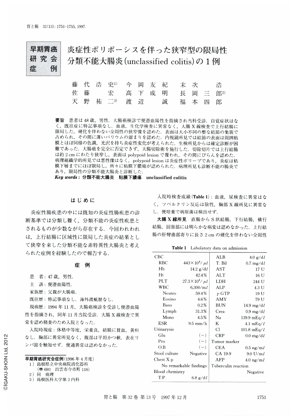
要旨 患者は48歳,男性.大腸癌検診で便潜血陽性を指摘され当科受診.自覚症状はなく,既往症に特記事項なし.血液,生化学検査に異常なく,大腸X線検査で上行結腸に限局した,硬化を伴わない全周性の狭窄像を認めた.表面は大小不同の整な結節の集簇で占められ,その間に薄いバリウムの溜まりを認めた.内視鏡所見では結節の表面は周囲粘膜とほぼ同様の色調,光沢を持ち炎症性変化が考えられた.生検所見からは確定診断が困難であった,大腸癌を完全に否定できず,大腸切除術を施行した.切除切片では上行結腸は約2cmにわたり狭窄し,表面はpolypoid lesionで覆われ,その間にびらんを認めた.病理組織学的所見では悪性像はなく,polypoid lesionは炎症性ポリープであり,炎症は粘膜下層までにほぼ限局し,所々に粘膜下膿瘍が認められた.病理所見も診断不能の腸炎であり,限局性の分類不能大腸炎と診断した.
The patient was 48-year-old male. He was admitted to our hospital due to positive occult blood of the stool. He had had no complaint and no contributing episode in his past history. On double contrast barium enema examination, a circular stenotic lesion without stiffness was shown in the ascending colon. This lesion was accompanied by polypoid lesions and shallow barium accumulations were seen among these lesions. Colonfiber examination showed that the surface of the polypoid lesions was almost of the same luster and color as that of the surrounding mucosa, thus indicating an inflammatory change. Although it was not histologically proved, there was a possibility of colon cancer, so right colectomy was performed. The ascending colon was stenosed for about 2 cm in length, where polypoid lesions with several erosions could be observed. Histologically no malignancy was observed. Polypoid lesions were composed of inflammatory polyps and inflammatory change was limited within the submucosal layer, and submucosal abscesses existed. Thus, unclassified colitis was our diagnosis.

Copyright © 1997, Igaku-Shoin Ltd. All rights reserved.


