Japanese
English
- 有料閲覧
- Abstract 文献概要
- 1ページ目 Look Inside
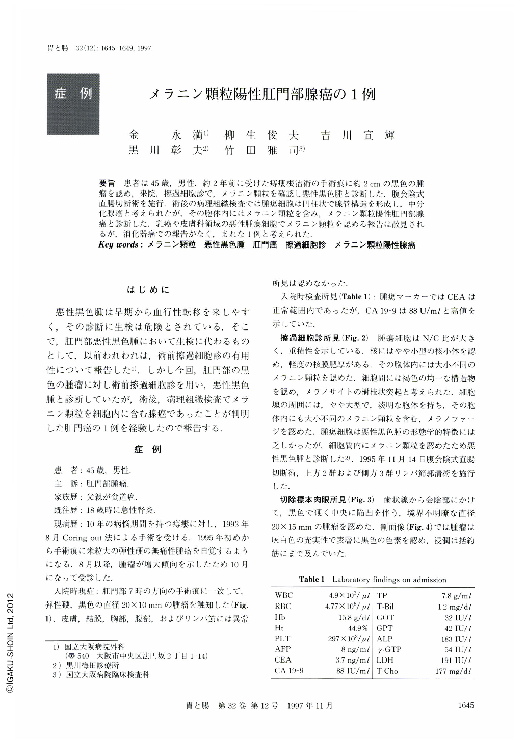
要旨 患者は45歳,男性.約2年前に受けた痔瘻根治術の手術痕に約2cmの黒色の腫瘤を認め,来院.擦過細胞診で,メラニン顆粒を確認し悪性黒色腫と診断した.腹会陰式直腸切断術を施行.術後の病理組織検査では腫瘍細胞は円柱状で腺管構造を形成し,中分化腺癌と考えられたが,その胞体内にはメラニン顆粒を含み,メラニン顆粒陽性肛門部腺癌と診断した.乳癌や皮膚科領域の悪性腫瘍細胞でメラニン顆粒を認める報告は散見されるが,消化器癌での報告がなく,まれな1例と考えられた.
The patient was a 45-year-old man. A tumor arose on the operation scar for anal fistula about two years ago. It was pigmented and about 2 cm in diameter. Touch smear cytology showed melanin granules in malignant cells. It was diagnosed as malignant melanoma and abdomino-perineal resection was performed. The final diagnosis obtained by histological examination and immunohistochemical staining revealed moderately differentiated adenocarcinoma which contained melanin granules. Since there was no report about pigmented adenocarcinoma of the digestive tract, we decided to report our case.

Copyright © 1997, Igaku-Shoin Ltd. All rights reserved.


