Japanese
English
- 有料閲覧
- Abstract 文献概要
- 1ページ目 Look Inside
- サイト内被引用 Cited by
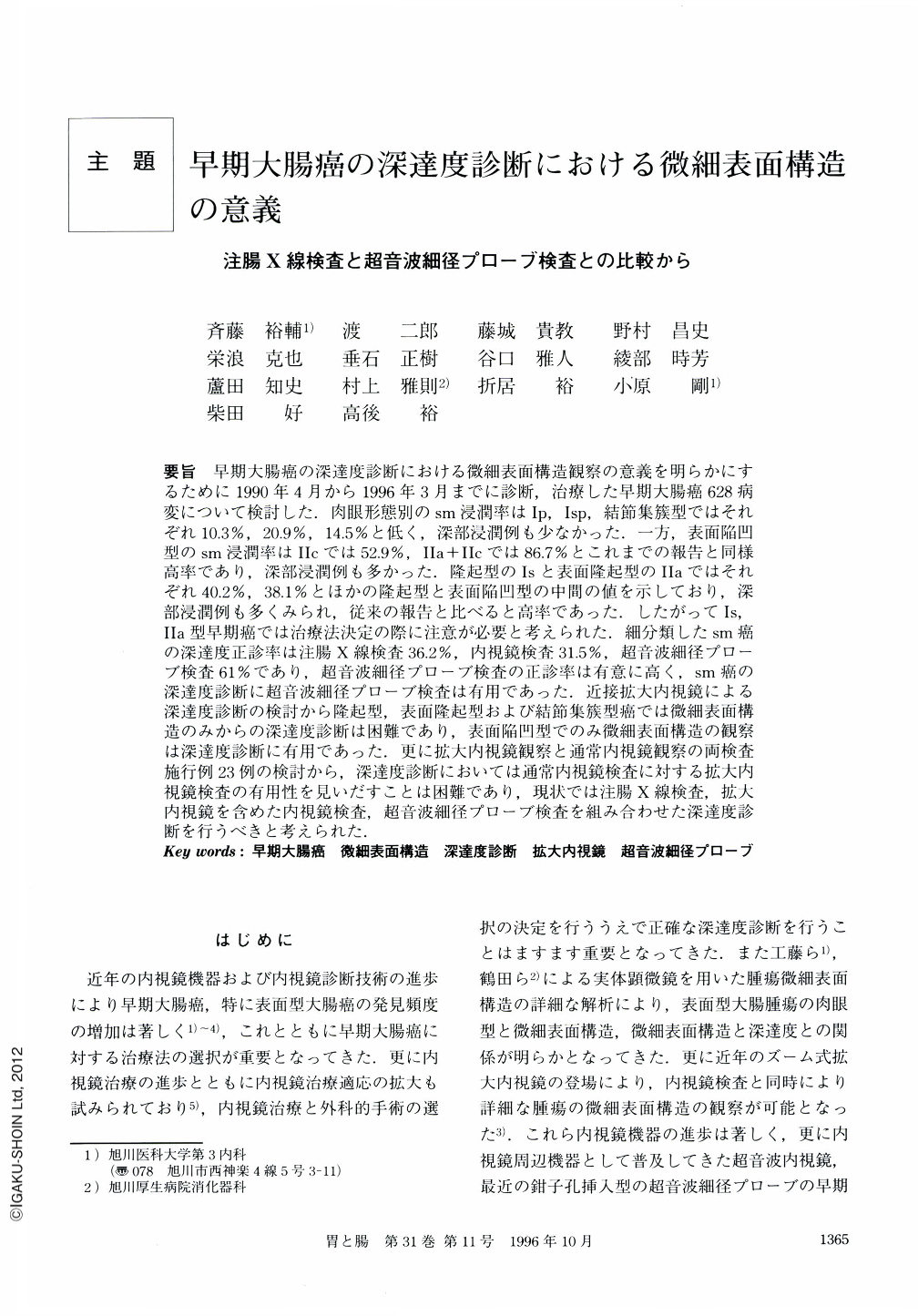
要旨 早期大腸癌の深達度診断における微細表面構造観察の意義を明らかにするために1990年4月から1996年3月までに診断,治療した早期大腸癌628病変について検討した.肉眼形態別のsm浸潤率はⅠp,Ⅰsp,結節集簇型ではそれぞれ10.3%,20.9%,14.5%と低く,深部浸潤例も少なかった.一方,表面陥凹型のsm浸潤率はⅡcでは52.9%,Ⅱa+Ⅱcでは86.7%とこれまでの報告と同様高率であり,深部浸潤例も多かった.隆起型のⅠsと表面隆起型のⅡaではそれぞれ40.2%,38.1%とほかの隆起型と表面陥凹型の中間の値を示しており,深部浸潤例も多くみられ,従来の報告と比べると高率であった.したがってⅠs,Ⅱa型早期癌では治療法決定の際に注意が必要と考えられた.細分類したsm癌の深達度正診率は注腸X線検査36.2%,内視鏡検査31.5%,超音波細径プローブ検査61%であり,超音波細径プローブ検査の正診率は有意に高く,sm癌の深達度診断に超音波細径プローブ検査は有用であった.近接拡大内視鏡による深達度診断の検討から隆起型,表面隆起型および結節集簇型癌では微細表面構造のみからの深達度診断は困難であり,表面陥凹型でのみ微細表面構造の観察は深達度診断に有用であった.更に拡大内視鏡観察と通常内視鏡観察の両検査施行例23例の検討から,深達度診断においては通常内視鏡検査に対する拡大内視鏡検査の有用性を見いだすことは困難であり,現状では注腸X線検査,拡大内視鏡を含めた内視鏡検査,超音波細径プローブ検査を組み合わせた深達度診断を行うべきと考えられた.
During the period between April of 1990 and March of 1996, 628 lesions of early colorectal carcinoma were studied to clarify the importance of the detailed surface structures for the diagnosis of depth of invasion in the early colorectal carcinoma. Submucosal invasion rates of the protruded type Ⅰp (pedunculated), Ⅰsp (subpedunculatred) and flat elevated nodule aggregating type were 10.3%, 20.9%,14.5%, respectively, submucosal massive invasion was infrequent. Submucosal invasion rates were high in the slightly depressed type (Ⅱc, 52.9%) and flat elevated with depression type (Ⅱa + Ⅱc, 86.7%) and many cases had massive invasion. Those results were consistent with previously reported invasion rates. In the protruded type Is and flat elevated type Ⅱa, Submucosal invasion rates were 40.2% and 38.1%, respectively. Therefore, we should be deliberate in diagnosing the depth of invasion of the protruded type Ⅰs and flat elevated type Ⅱa that were generally thought to be less aggressive than the types Ⅱc, and Ⅱa + Ⅱc. The diagnostic accuracy rates of depth of invasion of submucosal carcinoma by the detailed classification (sm1, sm2, sm3) were 36.2% in barium enema,31.5% in colonoscopic examination, and 61% in high-frequency ultrasound probe (HFUP). Thus, HFSP was an useful imaging technique in determining the depth of invasion of the submucosal involvement. In all protruded type, flat elevated type, and flat elevated nodule aggregating type, diagnosis of depth of invasion could not be correctly made only by detailed surface structure assessed by colonoscopic examination. On the other hand, in the types Ⅱc and Ⅱa + Ⅱc, assessment of detailed surface structure of the depressed floor was useful for diagnosis of depth of invasion. Moreover, there was no significant difference in the diagnostic accuracy rate of depth of invasion between magnified colonoscopic examination and conventional colonoscopic examination. In conclusion, the diagnosis of depth of invasion should be made by the combination of X-ray examination, colonoscopic examination (including magnified colonoscopic examination) and HFUP.

Copyright © 1996, Igaku-Shoin Ltd. All rights reserved.


