Japanese
English
- 有料閲覧
- Abstract 文献概要
- 1ページ目 Look Inside
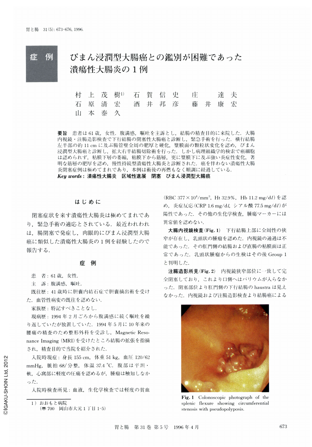
要旨 患者は61歳,女性.腹満感,嘔吐を主訴とし,結腸の精査目的に来院した.大腸内視鏡・注腸造影検査で下行結腸の閉塞性大腸癌と診断し,緊急手術を行った.横行結腸左半部の約11cmに及ぶ腸管壁全周の肥厚と硬化,漿膜面の顆粒状変化を認め,びまん浸潤型大腸癌と診断し,拡大右半結腸切除術を行った.しかし病理組織学的検索で癌細胞は認められず,粘膜下層の萎縮,粘膜下から筋層,更に漿膜下に及ぶ強い炎症性変化,著明な筋層の肥厚を認め,慢性持続型潰瘍性大腸炎と診断された.癌を伴わない潰瘍性大腸炎閉塞症例は極めてまれであり,本例は術後の再燃もなく順調に経過している.
A 61-year-old woman was admitted with the complaint of abdominal fullness and nausea. Colonofiber examination and a barium enema examination revealed an obstruction at at the upper descending colon. The biopsy was negative for carcinoma, but we performed laparotomy because of our diagnosis of ileus due to primary cancer of the descending colon. At her operation, the distal transverse colon revealed a long narrow lesion approximately 11 cm in length, giving the impression of diffusely infiltrating carcinoma. Because of this right hemicolectomy was performed with extensive lymph node dissection. In the narrow lesion of the transverse colon, there was high-grade annular stenosis and a markedly thickened wall. However, after pathological examination, there was no evidence of neoplasm. The lesion demonstrated atrophic mucosa, a markedly thickened muscle, large numbers of leukocytes in the tunica submucosa and muscularis. Inflammatory pseudopolyps were present at the anal margin of the narrow lesion. These findings led to a diagnosis of ulcerative colitis. Obstruction due to ulcerative colitis is rare. In this case, the postoperative course was uneventful and there has been no sign of recurrence.

Copyright © 1996, Igaku-Shoin Ltd. All rights reserved.


