Japanese
English
- 有料閲覧
- Abstract 文献概要
- 1ページ目 Look Inside
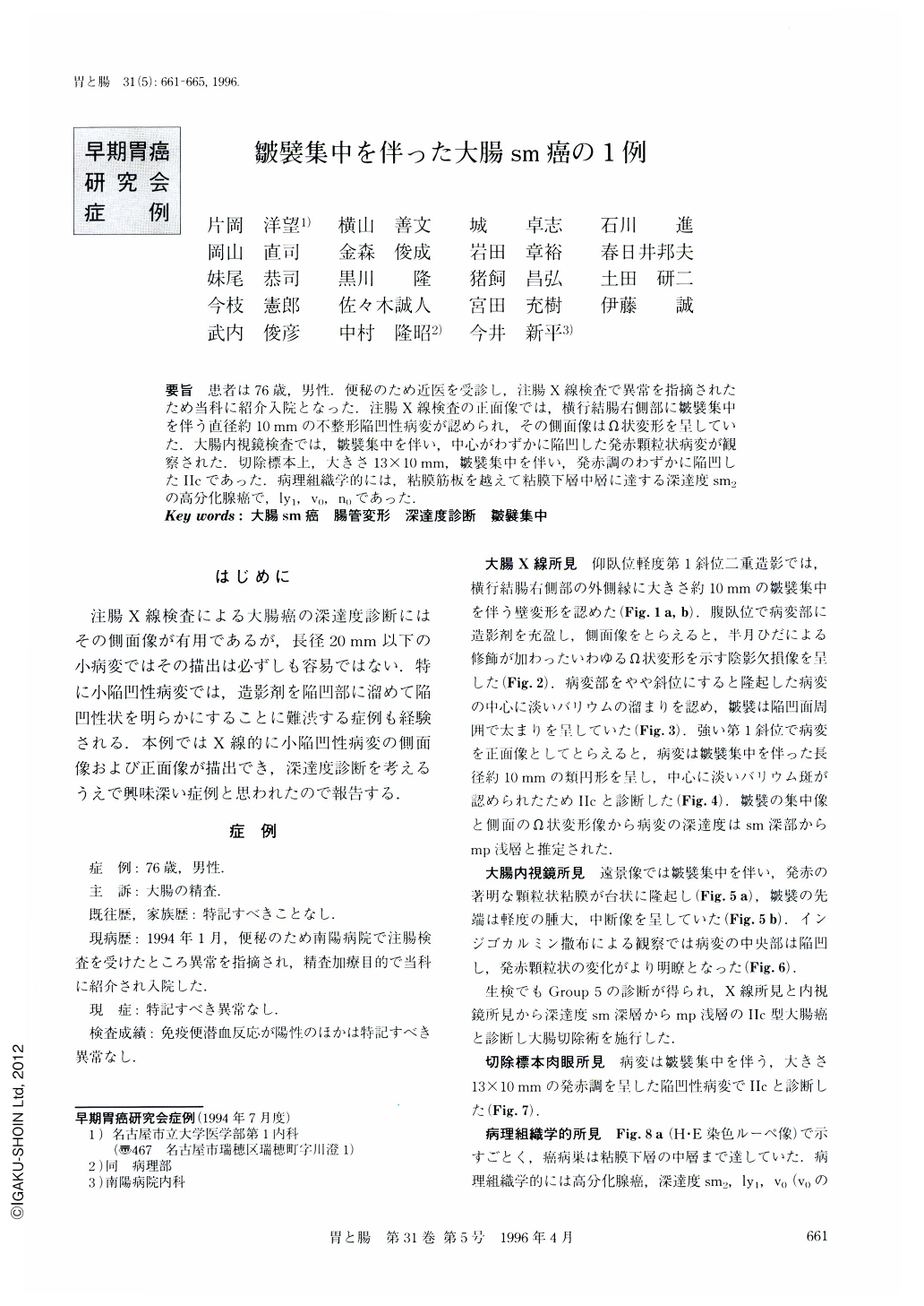
要旨 患者は76歳,男性.便秘のため近医を受診し,注腸X線検査で異常を指摘されたため当科に紹介入院となった.注腸X線検査の正面像では,横行結腸右側部に皺襞集中を伴う直径約10mmの不整形陥凹性病変が認められ,その側面像はΩ状変形を呈していた.大腸内視鏡検査では,皺襞集中を伴い,中心がわずかに陥凹した発赤顆粒状病変が観察された.切除標本上,大きさ13×10mm,皺襞集中を伴い,発赤調のわずかに陥凹したⅡcであった.病理組織学的には,粘膜筋板を越えて粘膜下層中層に達する深達度sm2の高分化腺癌で,ly1,v0,n0であった.
A 76-year-old man who had been found to have an abnormal lesion in the transverse colon at Nanyo hospital was admitted to our department. Barium enema showed a small irregularly-shaped slightly depressed lesion with converging folds in the transverse colon. The frontal view of barium enema study disclosed small barium spots in the center of the depression and the lateral view revealed an omega-shaped deformity. Colonoscopic examination showed an erythematous granular-surfaced lesion with converging folds. The resected specimen showed a reddish shallow depressed Ⅱc lesion, 13×10 mm in size. Histopathological examination disclosed well differentiated tubular adenocarcinoma with a slight adenomatous component, invading sm2. In this case, the omega-shaped deformity in the lateral view seemed to be important for evaluating the depth of the cancer invasion. In barium enema study, it is very important to disclose the depressed area and lateral deformity of the lesion for evaluating the depth of invasion of a small depressed colon cancer. Converging folds may be caused by thickning and fibrosis of mucosal muscle and submucosal cancer invasion with fibrosis.

Copyright © 1996, Igaku-Shoin Ltd. All rights reserved.


