Japanese
English
- 有料閲覧
- Abstract 文献概要
- 1ページ目 Look Inside
- サイト内被引用 Cited by
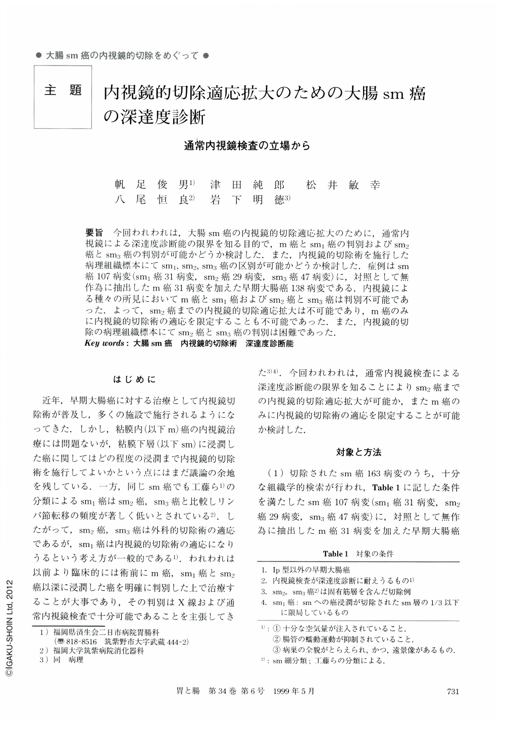
要旨 今回われわれは,大腸sm癌の内視鏡的切除適応拡大のために,通常内視鏡による深達度診断能の限界を知る目的で,m癌とsm1癌の判別およびsm2癌とsm3癌の判別が可能かどうか検討した.また,内視鏡的切除術を施行した病理組織標本にてsm1,sm2,sm3癌の区別が可能かどうか検討した.症例はsm癌107病変(sm1癌31病変,sm2癌29病変,sm3癌47病変)に,対照として無作為に抽出したm癌31病変を加えた早期大腸癌138病変である.内視鏡による種々の所見においてm癌とsm1癌およびsm2癌とsm3癌は判別不可能であった.よって,sm2癌までの内視鏡的切除適応拡大は不可能であり,m癌のみに内視鏡的切除術の適応を限定することも不可能であった.また,内視鏡的切除の病理組織標本にてsm2癌とsm3癌の判別は困難であった.
The aim of the present study is to examine the ability of endoscopic diagnosis to determine the depth of submucosal invasion in early colorectal cancer.
(1) Differentiation between mucosal (m) cancer and mildly submucosal invasive (sm1) cancer was undertaken using nine features identified endoscopically.
(2) Differentiation between moderately submucosal invasive (sm2) cancer and massively submucosal invasive (sm3) cancer was also undertaken using the same nine features identified endoscopicaly.
The nine endoscopic features used to assess and compare the groups were rigidity of the lumen arc, submucosal uniform elevation around the tumor, mucosal convergece, ulcer or erosion on the tumor, central depression, impression of tenseness, tumor redness, circumferetial white spots, and disappearance of glossy impression. (i) m and sm1 invasive cancer were not able to be differentiated by these endoscopic features. (ii) neither were sm2 invasive cancer and sm3 invasive cancer able to be differentiated by these endoscopic features.
From these results, it is concluded that indication for endoscopic resection is not able to be extended, by examining the clincal data reveald by the nine endoscopic features recorded in this paper.

Copyright © 1999, Igaku-Shoin Ltd. All rights reserved.


