Japanese
English
- 有料閲覧
- Abstract 文献概要
- 1ページ目 Look Inside
- サイト内被引用 Cited by
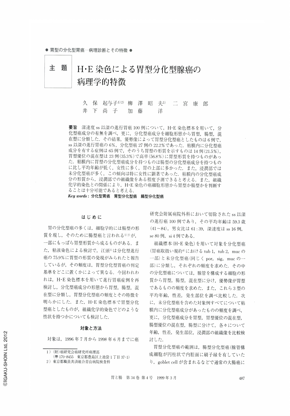
要旨 深達度ss以深の進行胃癌100例について,H・E染色標本を用いて,分化型癌成分の有無を調べ,更に,分化型癌成分を細胞形態から胃型,腸型,混在型に分類した.その結果,優勢像によって胃型分化型癌としたものは6例で,ss以深の進行胃癌の6%,分化型癌27例の22.2%であった.粘膜内に分化型癌成分を有する症例は65例で,そのうち胃型の形質を示すものは14例(21.5%),胃型優位の混在型は23例(35.3%)で高率(56.8%)に胃型形質を持つものがあった.粘膜内に胃型の分化型癌成分を持つものは腸型の分化型癌成分を持つものに比し平均年齢が低く,女性に多く,胃の上部に多かった.また,浸潤部では未分化型癌が多く,この傾向は特に女性に顕著であった.粘膜内の分化型癌成分の形質から,浸潤部での組織像をある程度予測できると考える.また,組織化学的染色との関係により,H・E染色の癌細胞形態から胃型か腸型かを判断することは十分可能であると考える.
Gastric carcinoma has long been classified into one of either two main histological types ; differentiatedtype or undifferentiated-type by Nakamura and Sugano (intestinal type or diffuse type by Lauren & Jarvi). Since the former is closely related to intestinal metaplasia of the stomach and the tumor cells almost always show intestinal properties, it is called intestinal type. However, some differentiated-type carcinomas are composed of cells resembling foveolar epithelium or pyloric gland cells or comprising hobnail cells with variable amounts of mucin, and these are often called differentiated-type carcinoma with gastric phenotype (DCGP). Although the incidence and detailed features of DCGP have been reported several times, the exact incidence and natural history of the tumor is not well known. So, in the present study, we investigated 100 consecutive cases of surgically resected advanced carcinomas, estimated the frequency of DCGP, and considered its natural history compared with that of genuine intestinal type carcinoma. When the differentiated-type carcinoma is divided into four subtypes according to the relative amount of gastric or intestinal phenotype, i.e gastric-type (G-type), gastric-type dominant mixed type (Gi-type), intestinal type dominant mixed type (Ⅰg-type) and intestinal type (Ⅰ-type), the number of G-type carcinoma was six, or 6% of the total number of advanced carcinomas and 22.2% of the total number (27) of differentiated-type carcinomas.
As for the differentiated-type carcinoma components in the mucosa of 65 cases, G-type, Gi-type, Ⅰg-type and Ⅰ-type were 14 (21.5%), 23 (35.3%), 15 (23.1%) and 10 (15.4%), respectively 〔3 (4.6%) were unclassified〕. The mean age of the cases with G-type carcinoma (56.1) was definitely younger than those for the cases with other types. The cases with G-type in the mucosa were female preponderant compared with the cases with Ⅰ-type, and tended to develop more frequently in the upper-third of the stomach. Further, this type changed more commonly to undifferentiated type during the course of invasion of the mucosa and/or the submucosa or deeper, compared with the cases with Ⅰ-type which usually keep the mucosal features of the differentiated type even after submucosal or ever deeper invasion. This tendency was more distinguished in females than in males. Thus, we think that the risk of differentiated-type carcinoma changing to undifferentiated type can be predicted even from the biopsy specimens, which are usually taken from superficial parts of the tumor.
Although recent progress in histochemistry has enabled us to differentiate the several properties of gastrointestinal epithelial cells, it has been shown that the discrimination of gastric carcinoma phenotype, gastric or intestinal, can be achieved more easily and exactly with H・E staining than with sophisticated histochemistry. Almost all the phenotypes decided on by H・E staining corresponded to the results obtained by histochemistry, and the latter did not always give us results definitive enough to enable discrimination of the gastric carcinoma phenotype.

Copyright © 1999, Igaku-Shoin Ltd. All rights reserved.


