Japanese
English
- 有料閲覧
- Abstract 文献概要
- 1ページ目 Look Inside
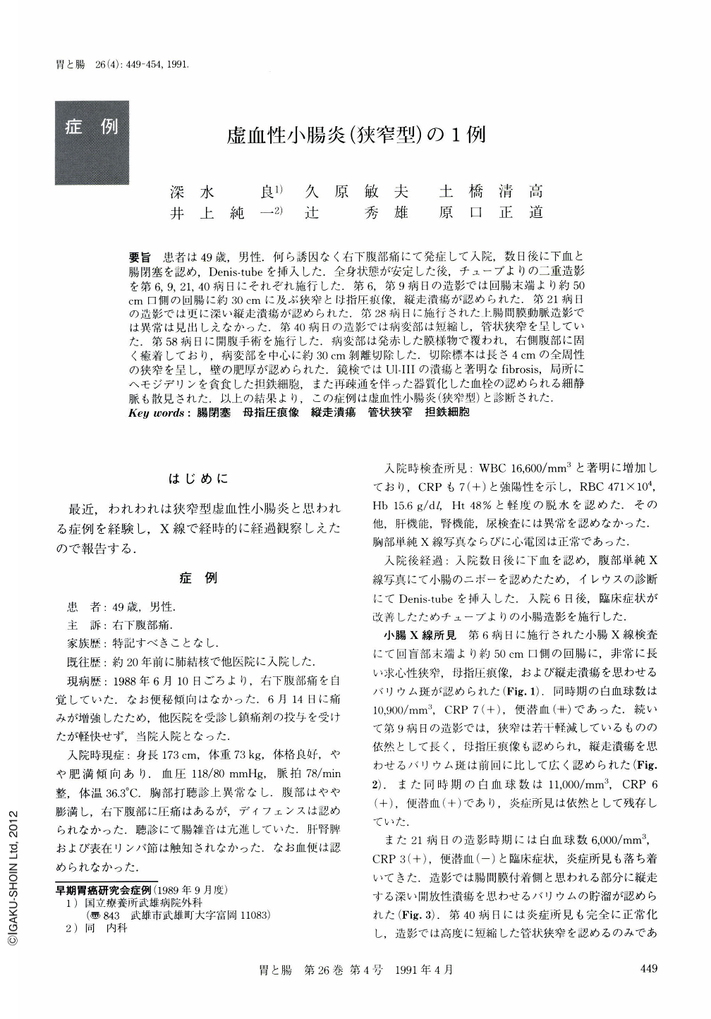
要旨 患者は49歳,男性.何ら誘因なく右下腹部痛にて発症して入院,数日後に下血と腸閉塞を認め,Denis-tubeを挿入した.全身状態が安定した後,チューブよりの二重造影を第6,9,21,40病日にそれぞれ施行した.第6,第9病日の造影では回腸末端より約50cm口側の回腸に約30cmに及ぶ狭窄と母指圧痕像,縦走潰瘍が認められた.第21病日の造影では更に深い縦走潰瘍が認められた.第28病日に施行された上腸間膜動脈造影では異常は見出しえなかった.第40病日の造影では病変部は短縮し,管状狭窄を呈していた.第58病日に開腹手術を施行した.病変部は発赤した膜様物で覆われ,右側腹部に固く癒着しており,病変部を中心に約30cm剝離切除した.切除標本は長さ4cmの全周性の狭窄を呈し,壁の肥厚が認められた.鏡検ではUⅠ-Ⅲの潰瘍と著明なfibrosis,局所にヘモジデリンを貪食した担鉄細胞,また再疎通を伴った器質化した血栓の認められる細静脈も散見された.以上の結果より,この症例は虚血性小腸炎(狭窄型)と診断された.
A 49-year-old male was admitted to our hospital because of right abdominal pain with no preceding symptoms. On the 9th hospital day contrast x-ray study using a Denis-tube showed a stricture involving about 30 cm of the terminal ileum. There were thumb-printing and longitudinal ulcer. On the 21st day the second contrast study showed a longitudinal ulcer which appeared to be deeper than before and a sclerotic lesion in the opposite wall. On the 28th day angiography of the superior mesenteric artery was performed showing no abnormality. On the 40th day the third contrast study showed the lesion of the decreased size and tubular narrowing. On the 58th day operation was performed. Approximately 30 cm of the ileum with the diseased portion at the center was resected. There was a very hard stricture of about 4 cm in length and a swelling of the wall.
Histological examination showed a UⅠ-Ⅲ ulcer, prominent fibrosis and hemosiderin devoured by the macrophages. Furthermore, there was an organized venous thrombosis with recanalization on the side of the serosa. Based on these results, this patient was diagnosed as having had ischemic enteritis (stricturing form).

Copyright © 1991, Igaku-Shoin Ltd. All rights reserved.


