Japanese
English
- 有料閲覧
- Abstract 文献概要
- 1ページ目 Look Inside
- 参考文献 Reference
- サイト内被引用 Cited by
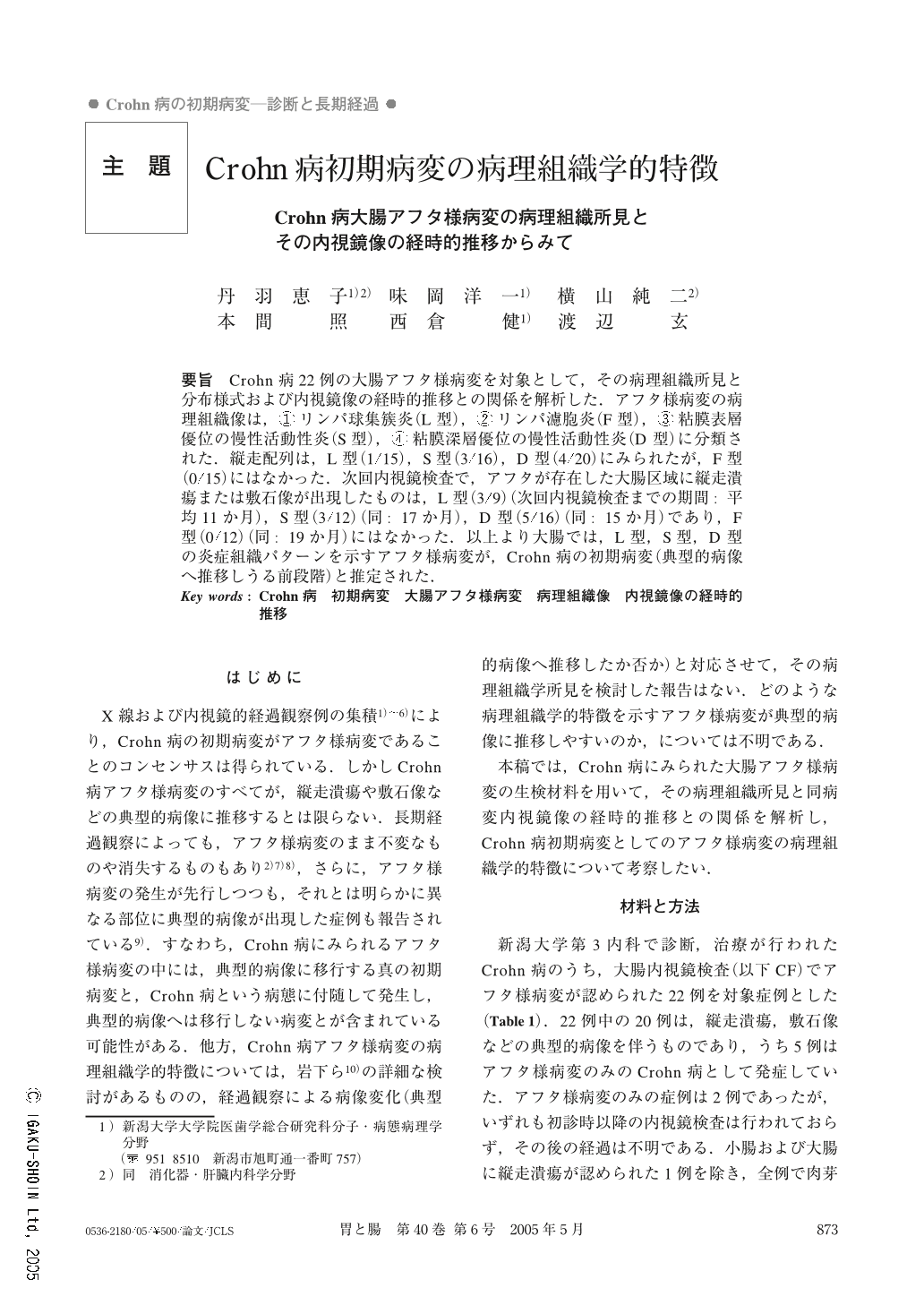
要旨 Crohn病22例の大腸アフタ様病変を対象として,その病理組織所見と分布様式および内視鏡像の経時的推移との関係を解析した.アフタ様病変の病理組織像は,①リンパ球集簇炎(L型),②リンパ濾胞炎(F型),③粘膜表層優位の慢性活動性炎(S型),④粘膜深層優位の慢性活動性炎(D型)に分類された.縦走配列は,L型(1/15),S型(3/16),D型(4/20)にみられたが,F型(0/15)にはなかった.次回内視鏡検査で,アフタが存在した大腸区域に縦走潰瘍または敷石像が出現したものは,L型(3/9)(次回内視鏡検査までの期間:平均11か月),S型(3/12)(同:17か月),D型(5/16)(同:15か月)であり,F型(0/12)(同:19か月)にはなかった.以上より大腸では,L型,S型,D型の炎症組織パターンを示すアフタ様病変が,Crohn病の初期病変(典型的病像へ推移しうる前段階)と推定された.
Histologic characteristics of aphthoid lesions of the colorectum and their distribution and transition as seen by endoscopic viewing were analyzed using 22 cases of Crohn's disease. Histologically, aphthoid lesions were grouped into 4 patterns : as ① Lymphocytic aggregation (type L), ② Lymph folliculitis (type F), ③ Chronic active inflammation mainly in the superficial mucosa (type S), and ④ Chronic active inflammation mainly in the deeper mucosa (type D). Longitudinal distribution was observed in type L (1/15), type S (3/16), and type D (4/20), but not in type F (0/15). Transition to longitudinal ulcer or cobblestone appearance at the next colonoscopic examination were seen in type L (3/9) (mean interval of 11 months), type S (3/12) (17 months), type D (5/16) (15 months), but not in type F (0/12) (19 months). These data suggest that aphthoid lesions of type L, S, and D patterns of inflammation would be the initial lesions of Crohn's disease of the colorectum.

Copyright © 2005, Igaku-Shoin Ltd. All rights reserved.


