Japanese
English
- 有料閲覧
- Abstract 文献概要
- 1ページ目 Look Inside
- 参考文献 Reference
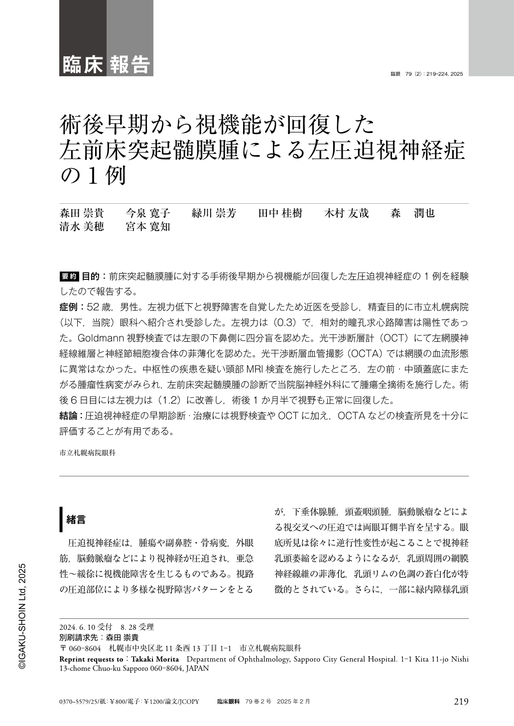
要約 目的:前床突起髄膜腫に対する手術後早期から視機能が回復した左圧迫視神経症の1例を経験したので報告する。
症例:52歳,男性。左視力低下と視野障害を自覚したため近医を受診し,精査目的に市立札幌病院(以下,当院)眼科へ紹介され受診した。左視力は(0.3)で,相対的瞳孔求心路障害は陽性であった。Goldmann視野検査では左眼の下鼻側に四分盲を認めた。光干渉断層計(OCT)にて左網膜神経線維層と神経節細胞複合体の菲薄化を認めた。光干渉断層血管撮影(OCTA)では網膜の血流形態に異常はなかった。中枢性の疾患を疑い頭部MRI検査を施行したところ,左の前・中頭蓋底にまたがる腫瘤性病変がみられ,左前床突起髄膜腫の診断で当院脳神経外科にて腫瘍全摘術を施行した。術後6日目には左視力は(1.2)に改善し,術後1か月半で視野も正常に回復した。
結論:圧迫視神経症の早期診断・治療には視野検査やOCTに加え,OCTAなどの検査所見を十分に評価することが有用である。
Abstract Purpose:We report a case of compressive optic neuropathy in which visual function recovered early after surgical excision of left anterior clinoid meningioma.
Case and Findings:A 52-year-old man was referred to our hospital with a chief complaint of decreased visual acuity and visual field defect in his left eye. His left visual acuity was 0.3 and relative afferent pupillary defect was observed. Optical coherence tomography(OCT)showed thinning of the retinal nerve fiber layer and ganglion cell complex in his left eye. Goldmann's visual field examination revealed quadrantanopia in the inferior nasal side of his left eye. We suspected a central nervous system disorder and performed head magnetic resonance imaging and found a mass lesion spanning the left anterior and middle skull bases. Left anterior clinoid meningioma was diagnosed and total tumor resection was performed. Postoperatively, visual acuity in the patient's left eye improved to 1.2 after 6 d and returned to baseline after 1.5 months.
Conclusions:For the early diagnosis and treatment of compression optic neuropathy, a comprehensive evaluation of test outcomes, including OCT angiography, must be conducted in conjunction with visual field examination and OCT.

Copyright © 2025, Igaku-Shoin Ltd. All rights reserved.


