Japanese
English
- 有料閲覧
PPV購入案内
1,320円 (1,200円+税10%) こちらは、489ページから493ページの文献です。購入には医書.jp本会員登録が必要です。
会員登録完了後に改めて上記「購入サイトへ」を選択してください。
または「購入サイトへ」選択後に「購入ログイン」、「新規会員登録」の順に進んでください。
会員登録について詳しくはをご確認ください。
- Abstract 文献概要
- 1ページ目 Look Inside
- 参考文献 Reference
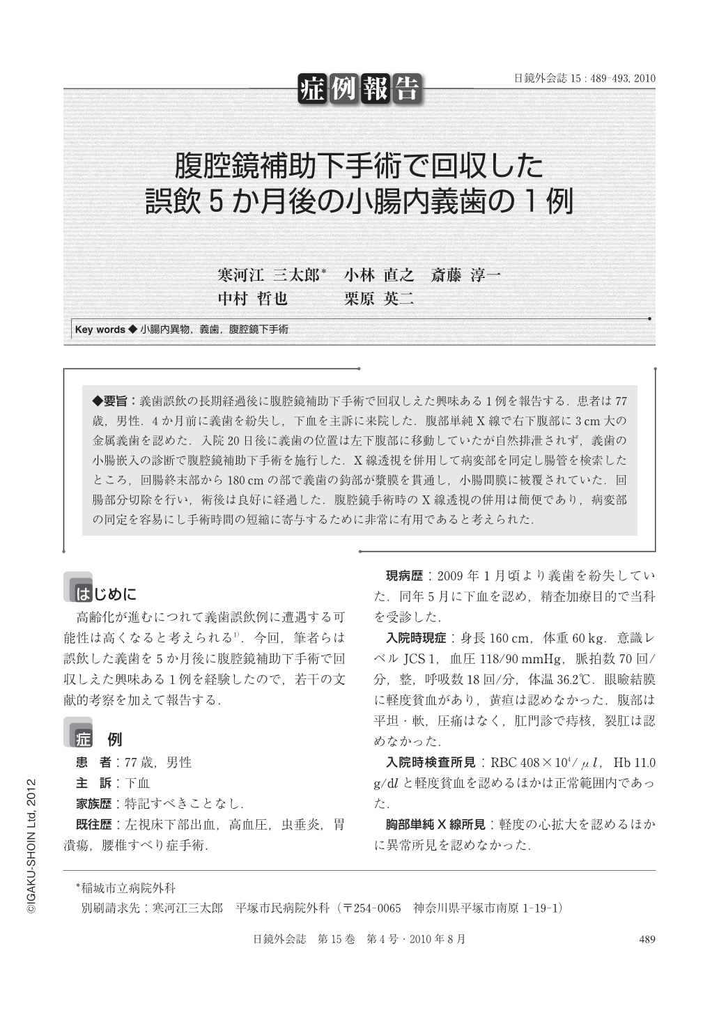
◆要旨:義歯誤飲の長期経過後に腹腔鏡補助下手術で回収しえた興味ある1例を報告する.患者は77歳,男性.4か月前に義歯を紛失し,下血を主訴に来院した.腹部単純X線で右下腹部に3cm大の金属義歯を認めた.入院20日後に義歯の位置は左下腹部に移動していたが自然排泄されず,義歯の小腸嵌入の診断で腹腔鏡補助下手術を施行した.X線透視を併用して病変部を同定し腸管を検索したところ,回腸終末部から180cmの部で義歯の鈎部が漿膜を貫通し,小腸間膜に被覆されていた.回腸部分切除を行い,術後は良好に経過した.腹腔鏡手術時のX線透視の併用は簡便であり,病変部の同定を容易にし手術時間の短縮に寄与するために非常に有用であると考えられた.
We describe a case in which laparoscopic surgery was effective for the removal of swallowed dental prosthesis 5 months after accidental swallowing. A 77-year-old male seen for melena was found on physical examination to have no symptom of peritonitis, and erect abdominal X-ray and computerized tomography showed a 3-cm-sized dental prosthesis in the small intestine. Twenty days later, abdominal X-ray showed the denture made a move to the left side of the pelvis but still never passed. Finally, laparoscopic surgery was performed under diagnosis of denture impaction in the intestinal wall. The wall of the ileum was perforated by clasp of the impacted denture, and the lesion was covered by the mesentery. The denture was removed by partially resecting the ileum. The postoperative course was fair. Laparoscopic surgery using serial radiography to confirm the presence and position of the swallowed denture is feasible and should be considered in cases in which conservative treatment is ineffective.
Copyright © 2010, JAPAN SOCIETY FOR ENDOSCOPIC SURGERY All rights reserved.