Japanese
English
- 有料閲覧
PPV購入案内
1,320円 (1,200円+税10%) こちらは、397ページから402ページの文献です。購入には医書.jp本会員登録が必要です。
会員登録完了後に改めて上記「購入サイトへ」を選択してください。
または「購入サイトへ」選択後に「購入ログイン」、「新規会員登録」の順に進んでください。
会員登録について詳しくはをご確認ください。
- Abstract 文献概要
- 1ページ目 Look Inside
- 参考文献 Reference
Ⅰ.はじめに
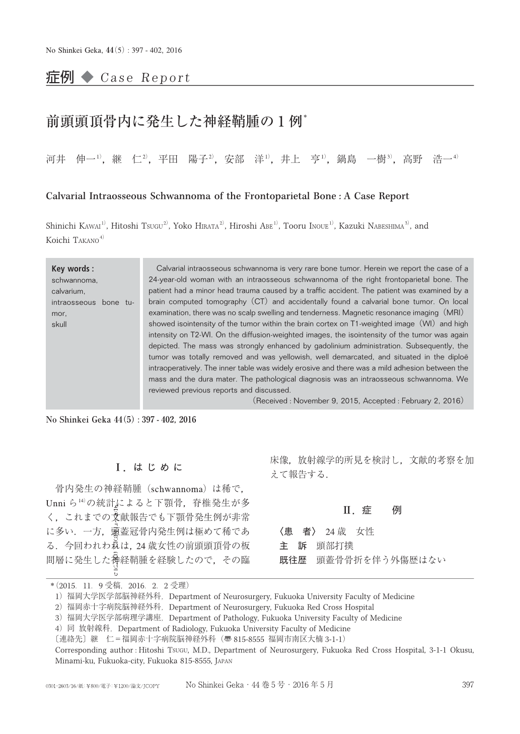
骨内発生の神経鞘腫(schwannoma)は稀で,Unniら14)の統計によると下顎骨,脊椎発生が多く,これまでの文献報告でも下顎骨発生例が非常に多い.一方,頭蓋冠骨内発生例は極めて稀である.今回われわれは,24歳女性の前頭頭頂骨の板間層に発生した神経鞘腫を経験したので,その臨床像,放射線学的所見を検討し,文献的考察を加えて報告する.
Calvarial intraosseous schwannoma is very rare bone tumor. Herein we report the case of a 24-year-old woman with an intraosseous schwannoma of the right frontoparietal bone. The patient had a minor head trauma caused by a traffic accident. The patient was examined by a brain computed tomography(CT)and accidentally found a calvarial bone tumor. On local examination, there was no scalp swelling and tenderness. Magnetic resonance imaging(MRI)showed isointensity of the tumor within the brain cortex on T1-weighted image(WI)and high intensity on T2-WI. On the diffusion-weighted images, the isointensity of the tumor was again depicted. The mass was strongly enhanced by gadolinium administration. Subsequently, the tumor was totally removed and was yellowish, well demarcated, and situated in the diploë intraoperatively. The inner table was widely erosive and there was a mild adhesion between the mass and the dura mater. The pathological diagnosis was an intraosseous schwannoma. We reviewed previous reports and discussed.
Copyright © 2016, Igaku-Shoin Ltd. All rights reserved.